01
Oxygen binds to the haem prosthetic group
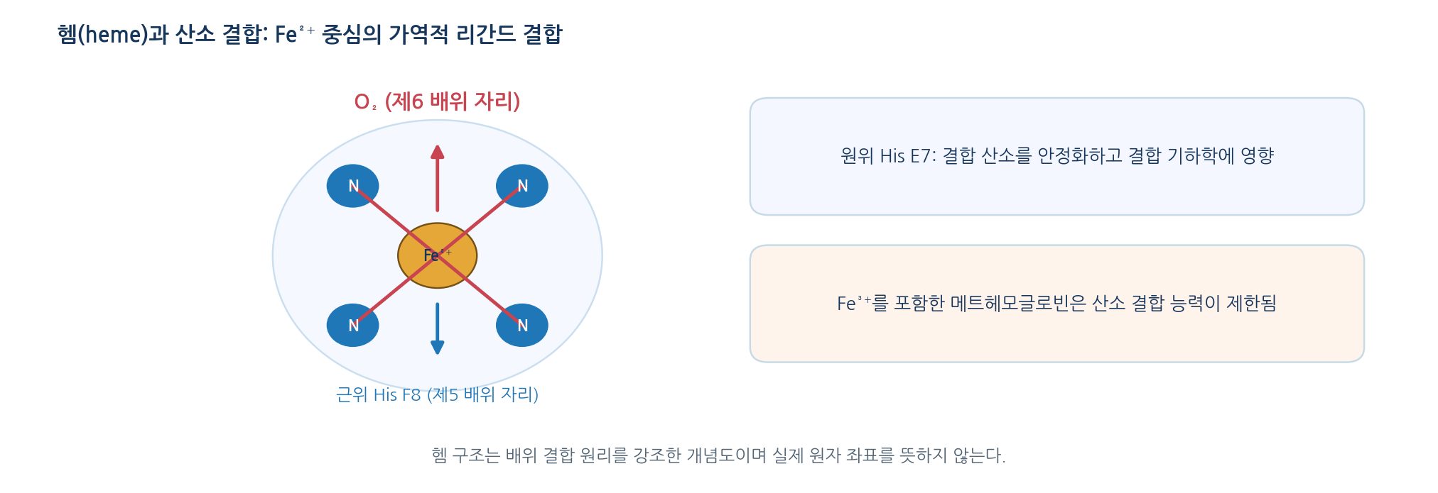
- Haem = porphyrin ring plus a central Fe; four nitrogens coordinate the iron
- Six coordination positions: four porphyrin N, the fifth from the proximal histidine (F8), the sixth is the O2 site
- Distal histidine (E7) fixes the orientation of bound oxygen and stabilises it
- Normal oxygen binding is reversible and requires Fe(II) haem
- Fe(III) methaemoglobin cannot transport oxygen; oxygenation is not the same as oxidation of the iron
02
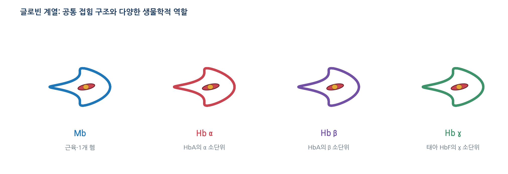
Globins form a family of oxygen-binding proteins
- The globin family shares an α-helical globin fold
- Myoglobin serves muscle; haemoglobin transports oxygen in erythrocytes
- Fetal HbF (α₂γ₂) differs in subunit composition from adult HbA (α₂β₂), so its binding properties differ
- Function is set by haem binding plus protein location, local conditions and inter-chain contacts
03
Myoglobin has a single oxygen-binding site
- One polypeptide with one haem gives one site, so no cooperativity is possible
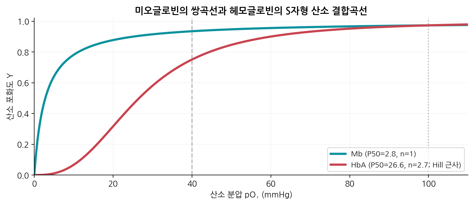
- Occupancy rises hyperbolically with oxygen partial pressure
- Typical physiological example: P50 about 2 to 3 mmHg, so occupancy is high at tissue pO2
- The protein therefore behaves as a storage and diffusion-facilitating protein
04
Protein–ligand binding can be described quantitatively
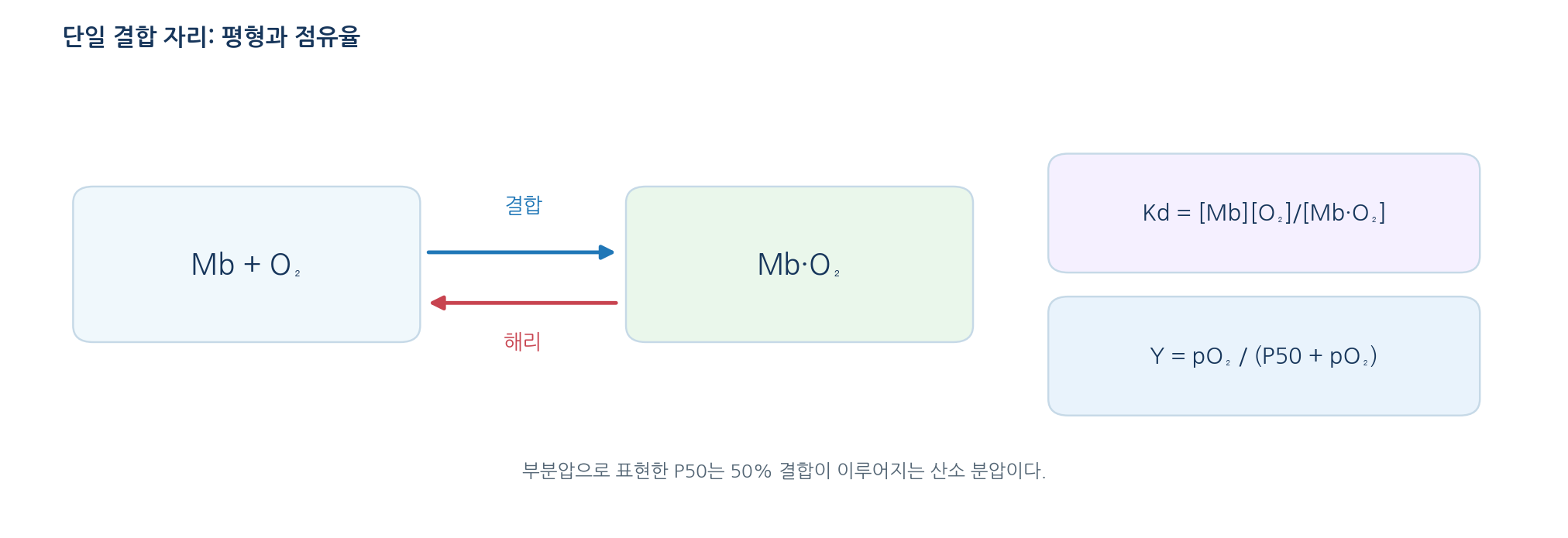
- A ligand is a molecule or ion that binds a protein selectively: Mb + O2 ⇌ Mb·O2
- Kd = [Mb][O2]/[Mb·O2]; a smaller Kd means higher occupancy at the same free concentration
- Occupancy Y = [Mb·O2]/([Mb] + [Mb·O2])
- Using partial pressure, Y = pO2/(P50 + pO2), where P50 is the half-saturation pressure
- A smaller P50 means higher affinity; Kd and P50 use different units and are not interchangeable
- Example: P50 2.8 mmHg at pO2 12 mmHg gives Y = 12/(2.8 + 12) ≈ 0.81
05
Protein structure determines how a ligand binds
- The haem iron alone does not explain the binding properties of a globin
- A hydrophobic pocket adjusts the water environment around haem and ligand
- Proximal and distal histidines set the coordination geometry and the direction of binding
- Access routes, steric hindrance and hydrogen bonding together determine selectivity and affinity
- Identical haem in different protein environments gives different relative reactivity toward O2 and CO
06
Haemoglobin transports oxygen in the blood
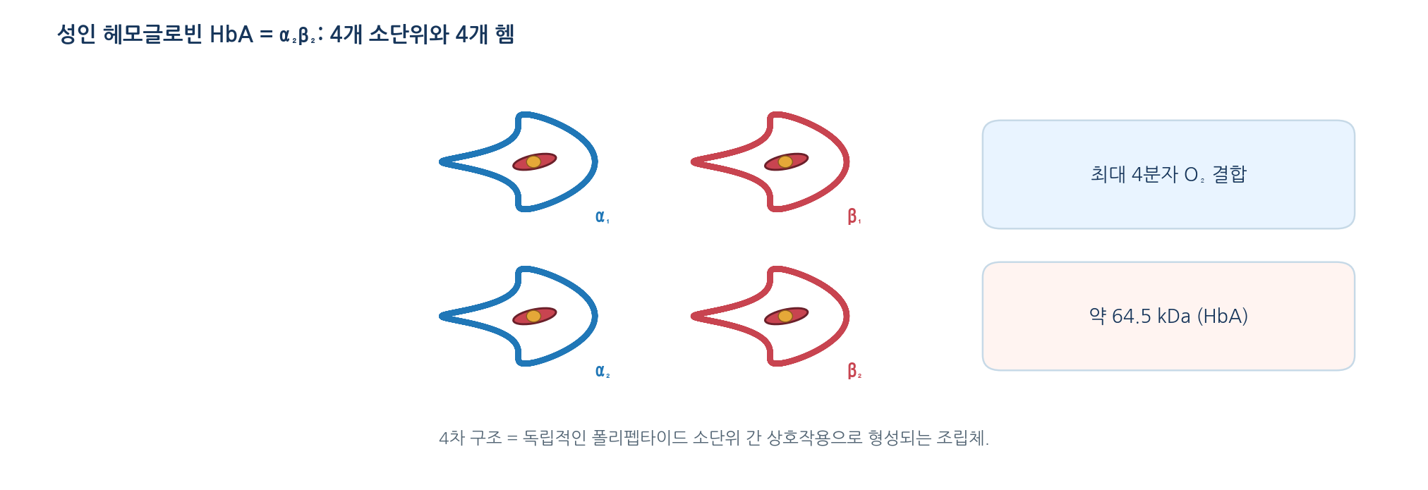
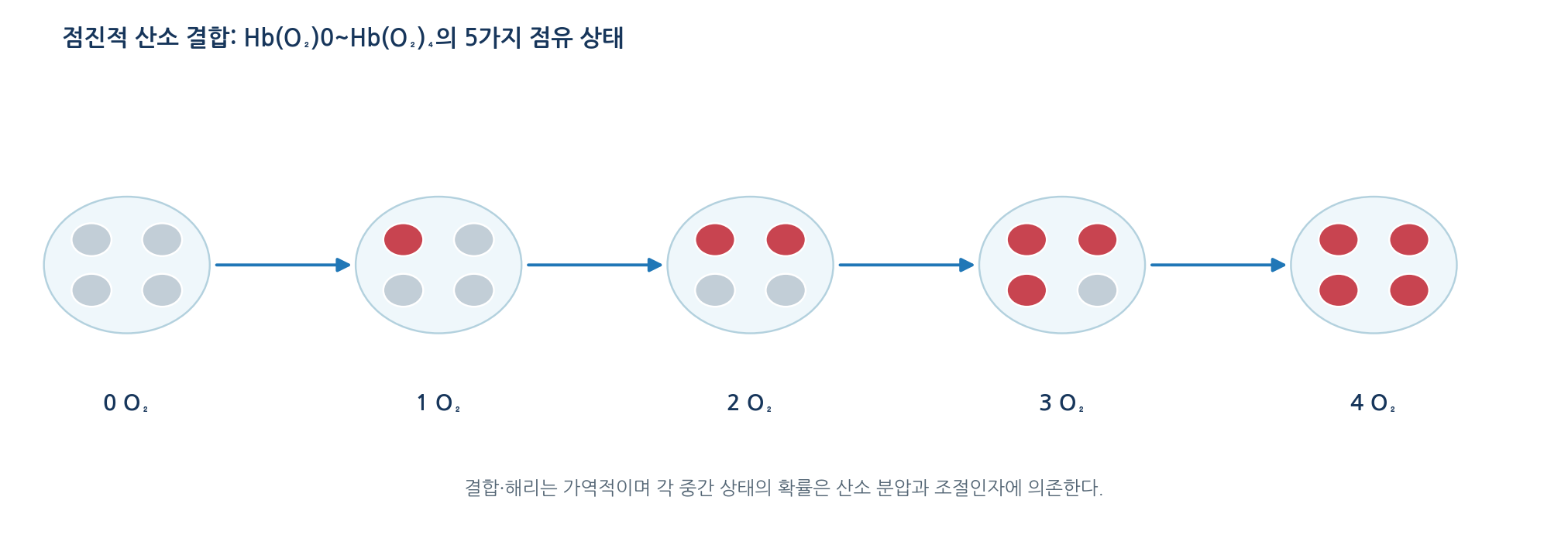
- HbA is an α₂β₂ tetramer with one haem per subunit, so up to four O2 per molecule
- Molecular mass about 64.5 kDa; most oxygen travels bound reversibly to Hb in erythrocytes
- Saturation is the fraction of haem sites filled; it differs from the total oxygen content of blood
- Dissolved oxygen in plasma is small; haemoglobin is the main carrier
07
Haemoglobin subunits resemble myoglobin structurally
- The α and β chains differ in sequence from Mb but share the globin fold
- Each chain carries one haem-binding pocket
- The difference is the quaternary structure formed by subunit contacts
- Those contact surfaces are the route by which binding at one haem affects the others
08
The structure of haemoglobin changes when oxygen binds
- O2 binding moves the iron and rearranges the proximal histidine and the F helix
- The subunit interface then changes and transmits the change to neighbouring chains
- T (tense) state has low affinity; R (relaxed) state has high affinity
- Real haemoglobin also occupies partially ligated states and several conformations, so T/R is a simplification
09
Haemoglobin binds oxygen cooperatively
- Positive cooperativity means binding at one site makes binding at the others more favourable
- The curve rises slowly at low pO2, steeply in the middle range and then saturates
- The sigmoid shape allows efficient loading in the lung and substantial unloading in tissue
- Without cooperativity, transport efficiency would fall substantially
10
Cooperative binding can also be described quantitatively
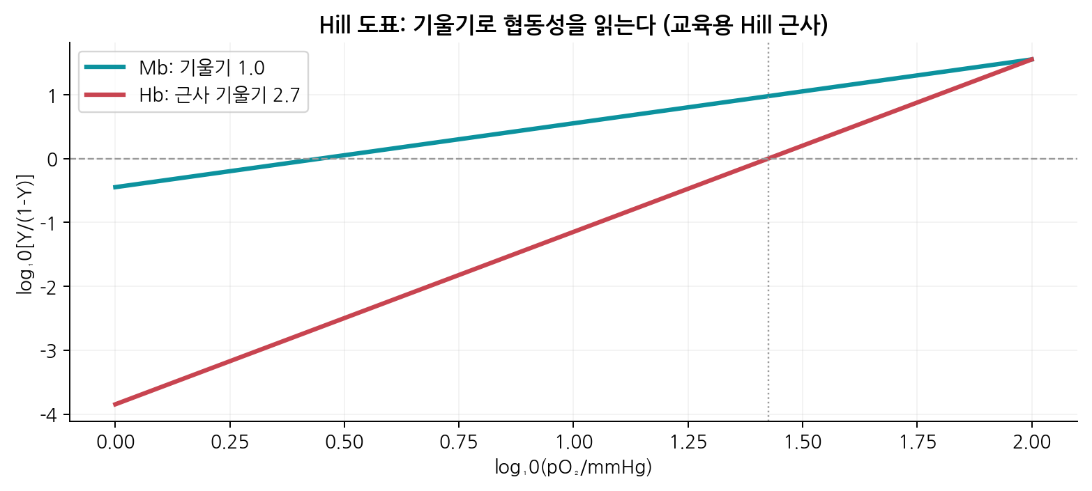
- Hill equation: Y = pO2^n/(P50^n + pO2^n), where n is the Hill coefficient
- For HbA in the mid-saturation range n ≈ 2.7, an index of positive cooperativity
- n is not the number of binding sites (there are four) and the equation is an empirical approximation
- Linearised form: log[Y/(1−Y)] = n·log pO2 − n·log P50, so the slope gives n
- The slope can vary between pO2 ranges, so the fitted interval must be stated
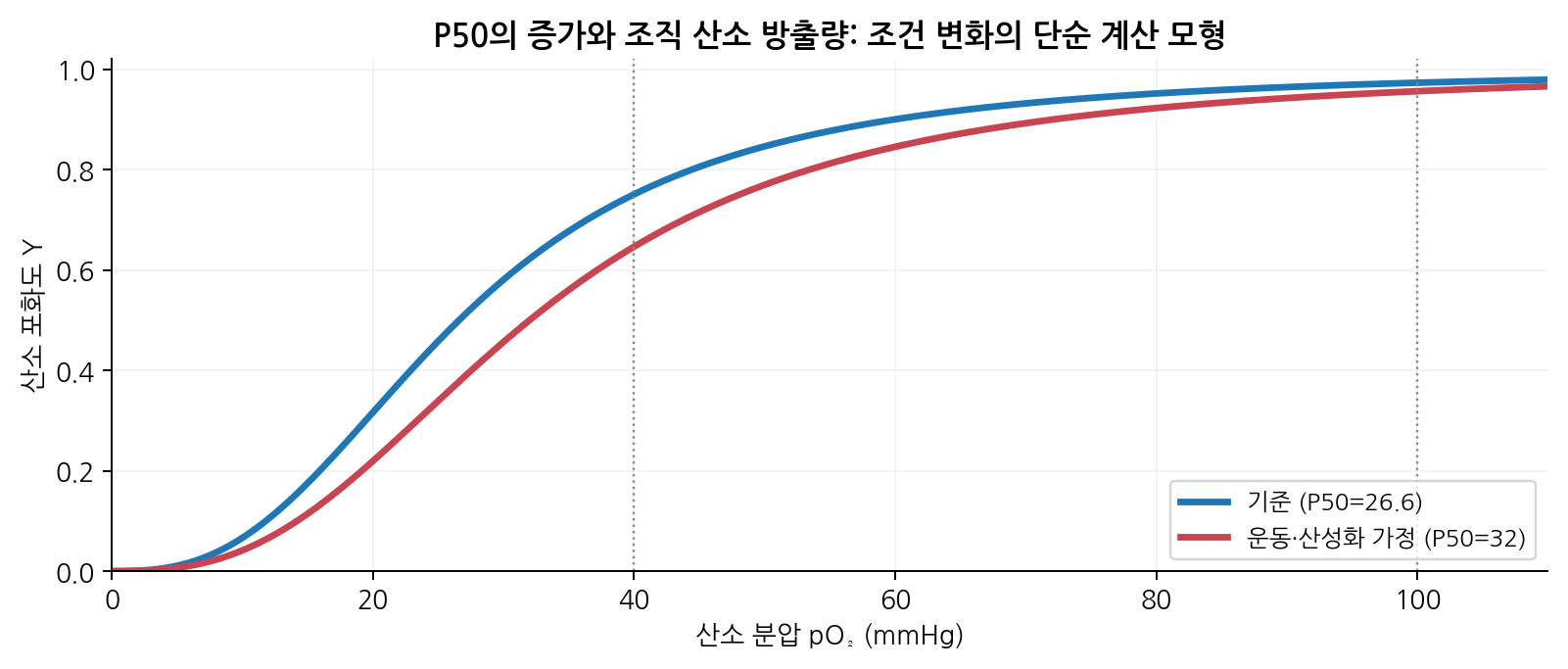
- Example: P50 26.6 mmHg, n 2.7, pO2 40 mmHg gives Y ≈ 0.750
11
Two classical models explain the mechanism of cooperativity
- MWC (concerted): subunits switch together between T and R, and ligand shifts the equilibrium
- KNF (sequential): each binding event changes structure and passes the change to neighbours
- Both are classical simplifications and neither matches the full structural dynamics
- The models differ in how far a conformational change is shared across subunits
12
[Separate note 5-1] Carbon monoxide: the quiet killer
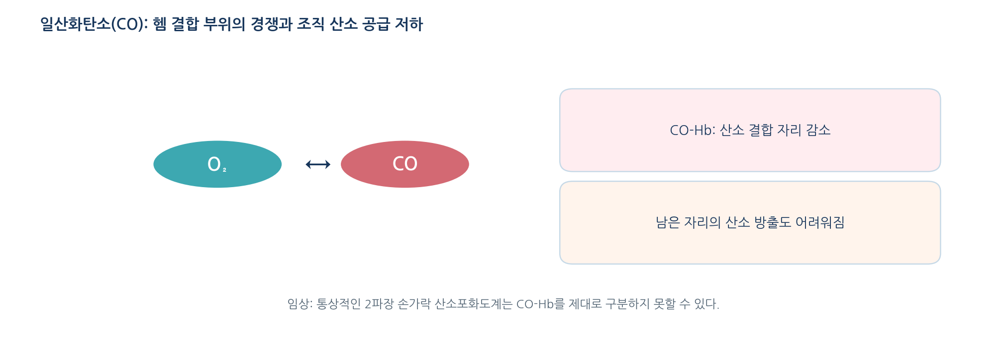
- CO is a colourless, odourless toxic gas that binds haem iron far more strongly than O2 (often cited as 200-fold or more)
- Two toxic routes: it occupies oxygen sites and it raises the affinity of the remaining sites, hindering release in tissue
- It is also toxic to cellular respiration
- A standard two-wavelength finger pulse oximeter cannot distinguish CO-Hb from O2-Hb and may miss the defect
- Move the casualty to safety and seek emergency care; diagnosis uses a CO-Hb measuring device
- Generating or inhaling CO in school experiments is never permitted
13
Haemoglobin also contributes to H⁺ and CO₂ transport
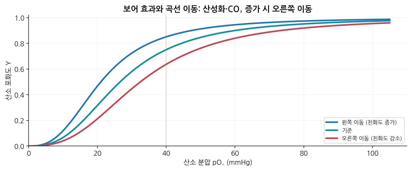
- Bohr effect: falling pH and rising CO2 lower oxygen affinity and shift the curve to the right
- This promotes release in tissue; in the lung, CO2 removal and higher pH favour binding
- Rising temperature also favours oxygen release
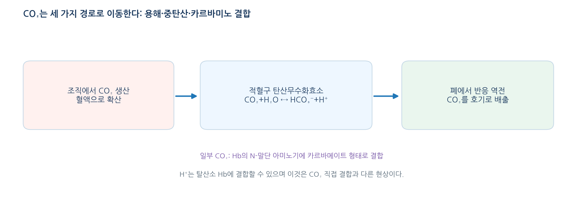
- CO2 travels in three forms: bicarbonate, dissolved CO2 and carbamino compounds on haemoglobin
- Carbonic anhydrase accelerates CO2 + H2O ⇌ HCO3− + H+
- CO2 does not bind the haem oxygen site; it reacts mainly with the N-terminal amino group of globin
14
2,3-BPG modulates oxygen binding by haemoglobin
- 2,3-BPG is a glycolytic metabolite of erythrocytes and carries negative charge
- It binds the central cavity of deoxyhaemoglobin, stabilising the T state and shifting the curve right
- The result is more oxygen release in tissue; it does not compete with O2 at the haem site
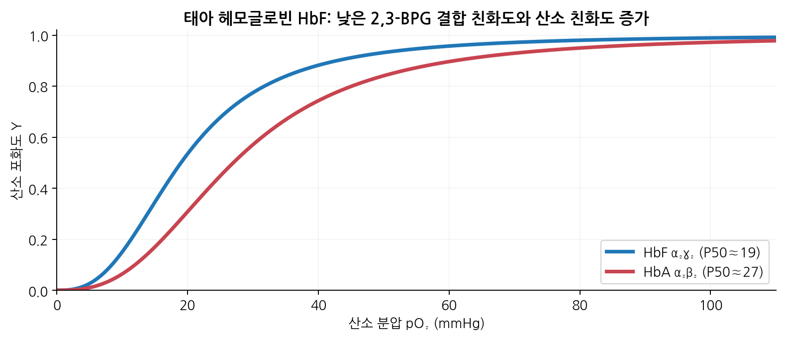
- HbF interacts only weakly with 2,3-BPG, so its affinity is higher (example P50 19 versus 27 mmHg)
- This favours transfer from mother to fetus; actual P50 varies with pH, temperature, CO2 and 2,3-BPG
15
Sickle cell disease is a molecular disease of haemoglobin
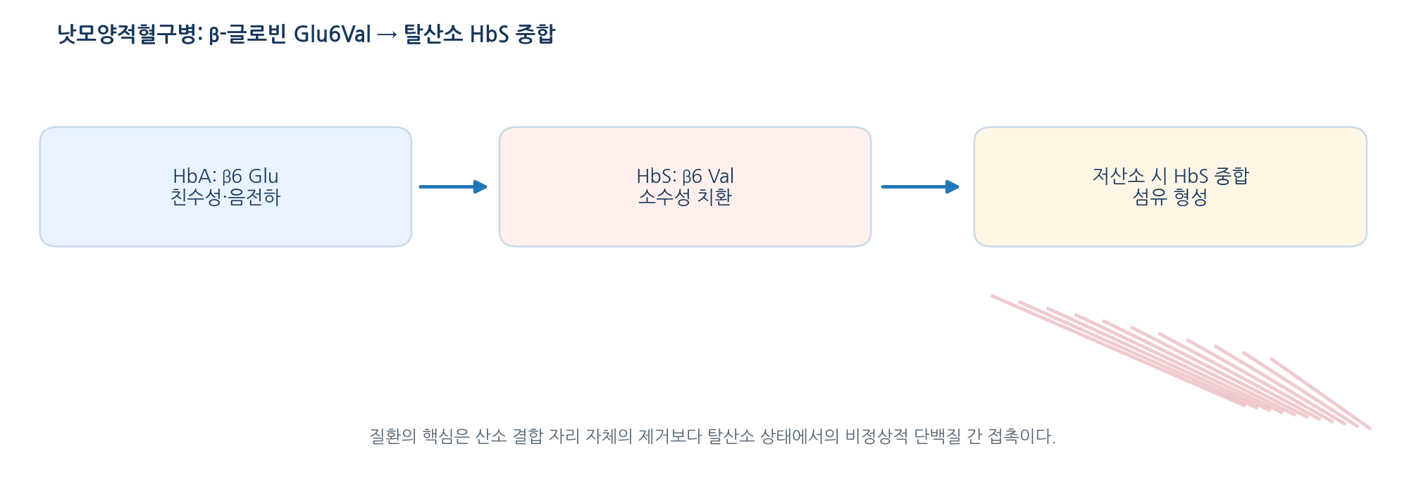
- A β-globin gene variant produces HbS; the classic change is β6 Glu to Val (written Glu7Val in some numbering)
- The hydrophobic Val lets deoxy HbS contact other HbS molecules, nucleating polymers and long fibres
- Consequences include loss of red-cell deformability, sickling, vaso-occlusion and haemolysis
- The key point is a surface change from one substitution, not the loss of the haem site
- Low oxygen promotes polymerisation through a molecular and cellular cascade
16
Oxygen delivery and release can be read numerically
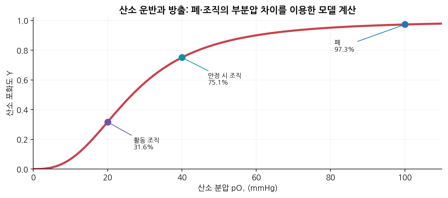
- With the teaching Hill approximation (P50 26.6 mmHg, n 2.7), lung pO2 100 gives about 97.3% saturation
- Resting tissue pO2 40 gives about 75.0%, and active tissue gives more release at lower pO2
- Rising temperature and CO2, and changes in pH and 2,3-BPG, can be modelled as a higher P50
- Actual delivery also depends on Hb concentration, blood flow and how fully capillary equilibration occurs
- A saturation difference alone does not explain the whole physiology of oxygen transport
References
| Protein | Typical composition | Main location and function | Cooperativity |
|---|---|---|---|
| Myoglobin (Mb) | One chain, one haem | Muscle: oxygen storage, facilitated diffusion | None |
| Adult HbA | α₂β₂, four haem groups | Erythrocytes: oxygen transport | Positive |
| Fetal HbF | α₂γ₂, four haem groups | Favours oxygen supply to the fetus | Positive |