01
Immune responses arise from cooperation between specialised cells and proteins
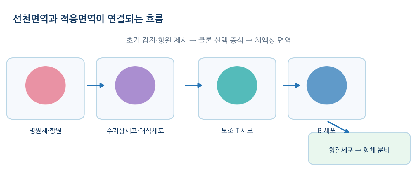
- Innate immunity uses skin and mucosal barriers, phagocytes, natural killer cells and complement for a rapid, low-specificity response
- Adaptive immunity selectively activates lymphocytes with diverse antigen receptors, giving high specificity and memory
- The two arms are linked by antigen presentation and cytokine signalling rather than separate systems
- Antigen = a molecule recognised by antibody or receptor; immunogen = a substance that actually elicits a response, so not every antigen is a strong immunogen
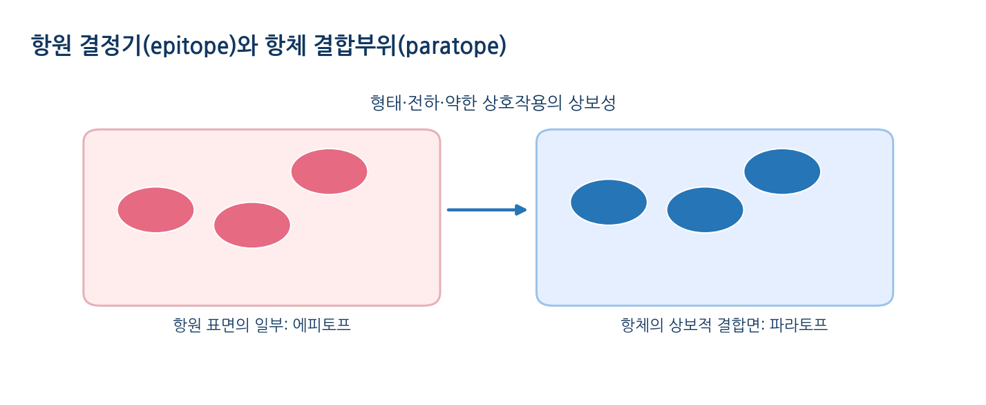
- Epitope = the local site recognised by an antibody, either a continuous sequence or a conformational patch
- Antibodies recognise the free antigen surface, whereas αβ T-cell receptors recognise peptide bound to MHC
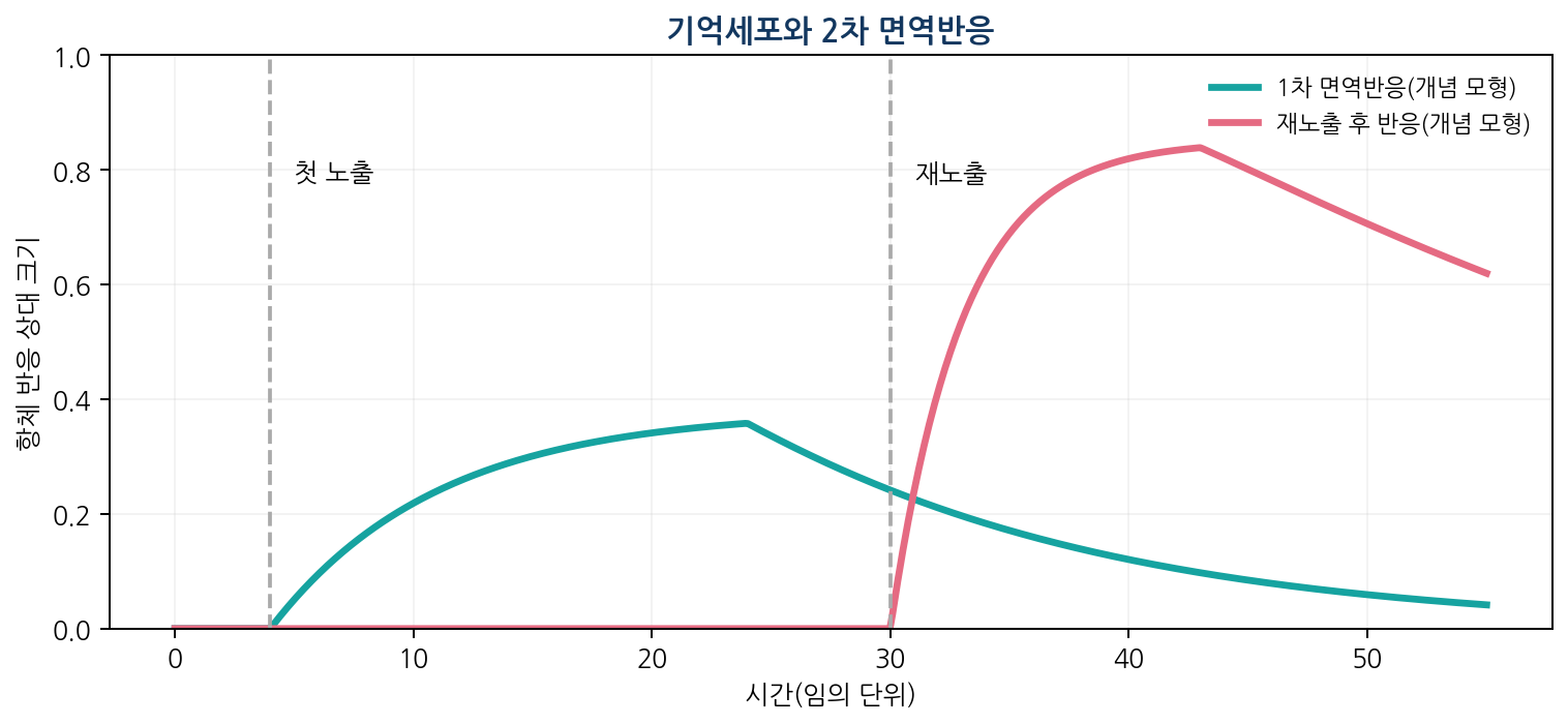
- B cells express a BCR after gene rearrangement, then clonally expand and differentiate into plasma and memory cells
- Somatic hypermutation, affinity maturation and class switching change effector function but do not themselves create new epitope recognition
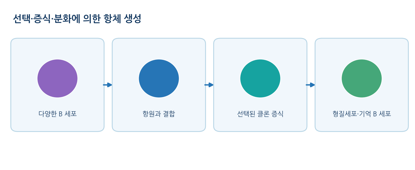
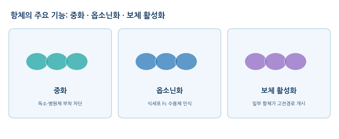
- Antibody actions: neutralisation, opsonisation, classical complement activation and Fc-receptor-mediated regulation
02
An antibody has two identical antigen-binding sites
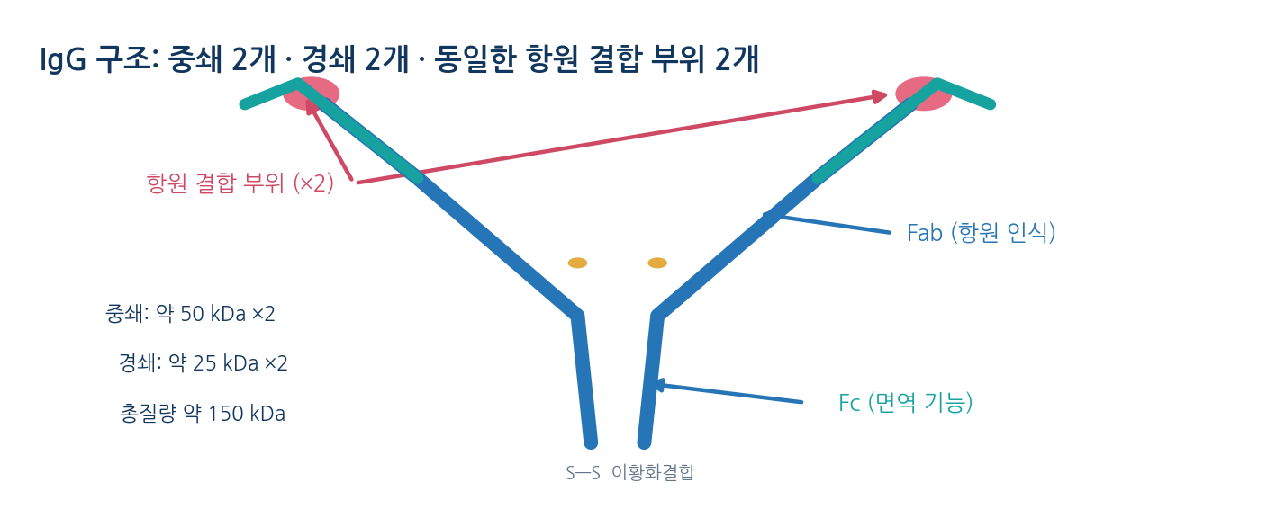
- An IgG monomer has two identical heavy chains (about 50 kDa) and two identical light chains (about 25 kDa), about 150 kDa in total
- Disulfide bonds between heavy chains and between heavy and light chains, plus non-covalent contacts, hold the fold together
- Paired V_H and V_L domains at the arm tips form one antigen-binding site each, so a monomer has two
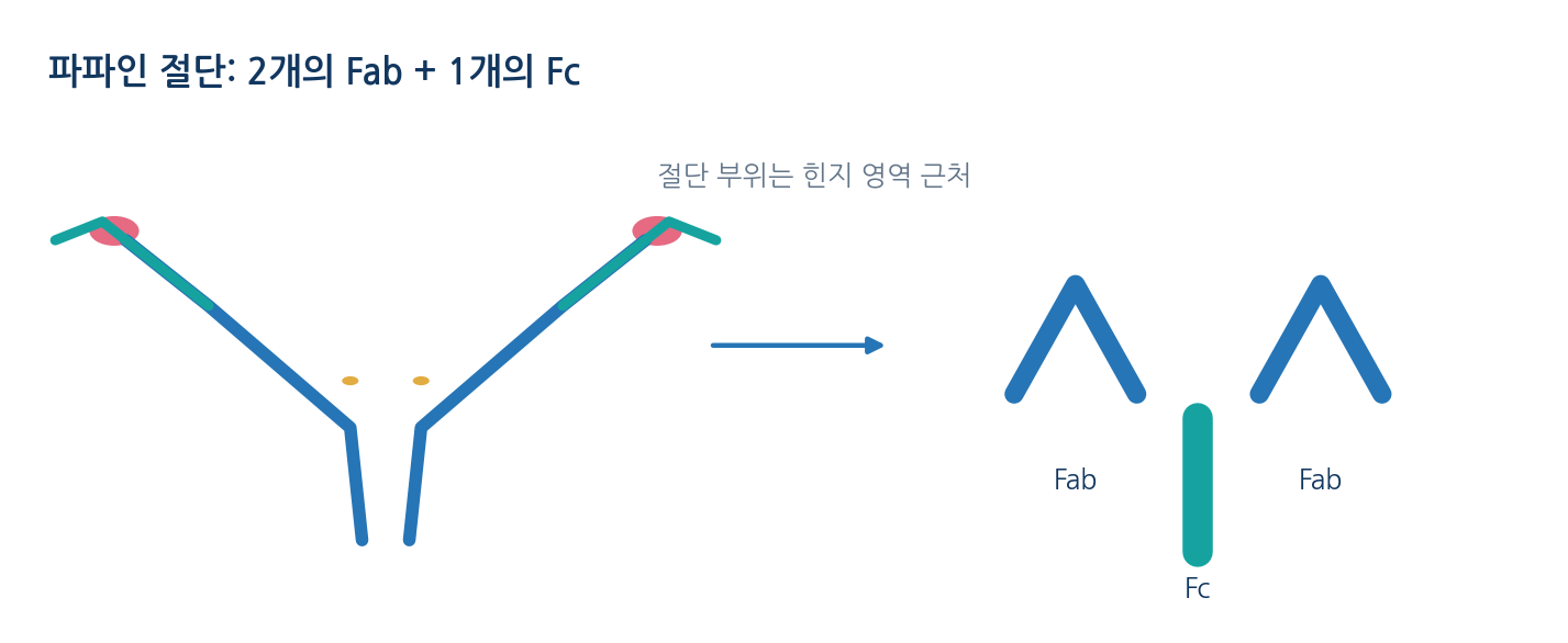
- Fab carries antigen recognition; Fc engages Fc receptors and some complement components to mediate effector function
- The hinge allows the arms to move; limited papain digestion yields two Fab fragments and one Fc fragment
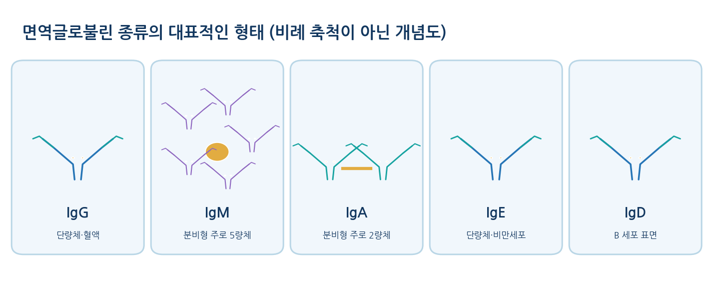
- Five isotypes (IgG, IgM, IgA, IgE, IgD) are defined by heavy-chain constant regions
- Secreted IgM is mostly pentameric and secreted IgA mostly dimeric, so the claim that every antibody is bivalent is inaccurate
03
Antibodies bind a specific antigen strongly and reversibly
- Selective recognition rests on partial complementarity of shape, charge distribution, polar groups and hydrophobic surface
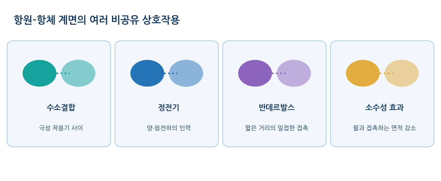
- Binding combines hydrogen bonds, electrostatic interactions, van der Waals forces and the hydrophobic effect, with no new covalent bond
- Rearrangement of the hydration layer and conformational change also contribute to the binding free energy
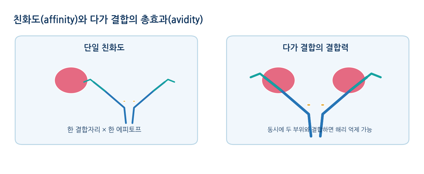
- Specificity = ability to distinguish targets; affinity = strength at one site with one epitope; avidity = combined multivalent stability
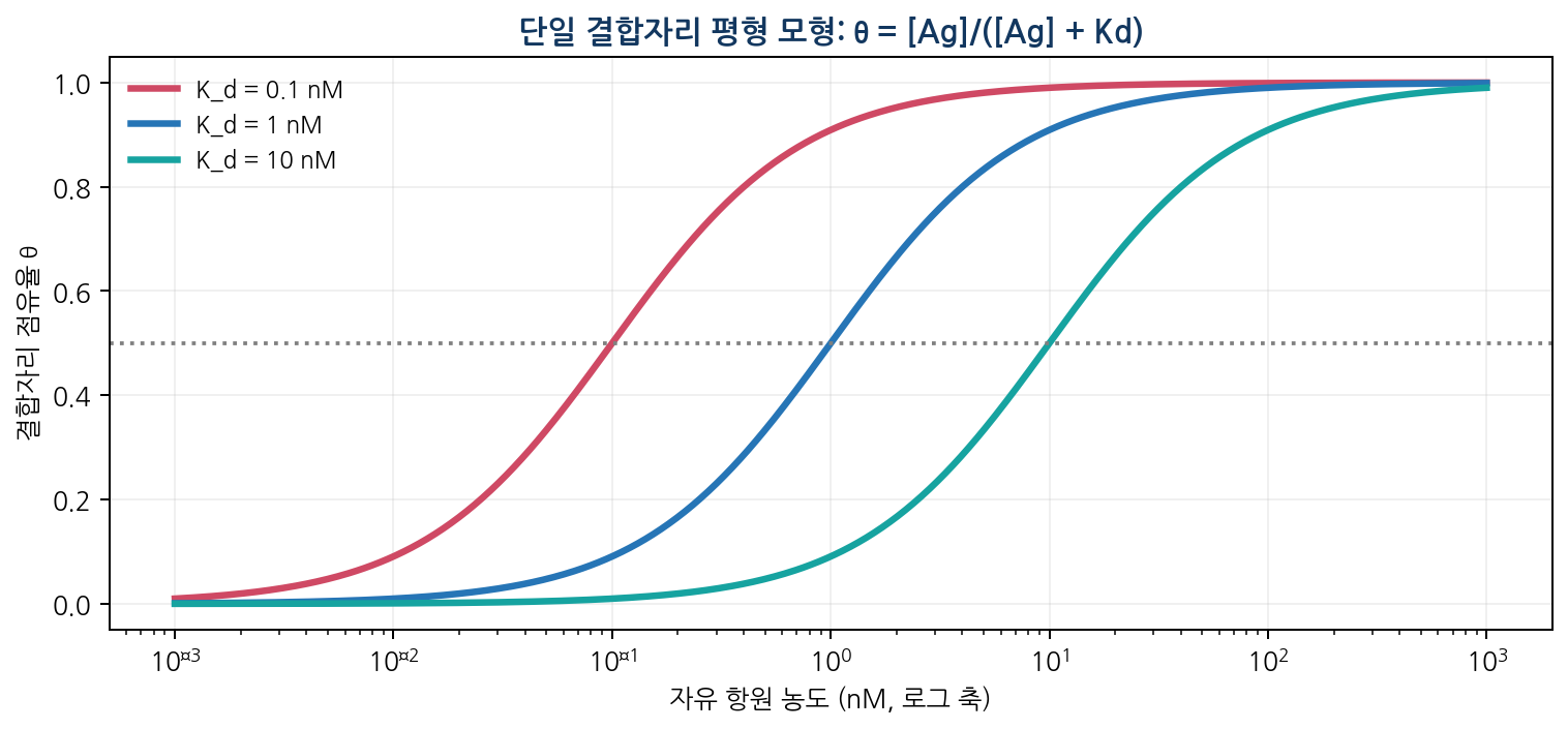
- Kd = [Ab][Ag]/[Ab·Ag] = koff/kon, and θ = [Ag]/(Kd + [Ag]), so θ = 0.5 when [Ag] = Kd
- Example: Kd 2 nM with 6 nM free antigen gives θ = 6/(2 + 6) = 0.75
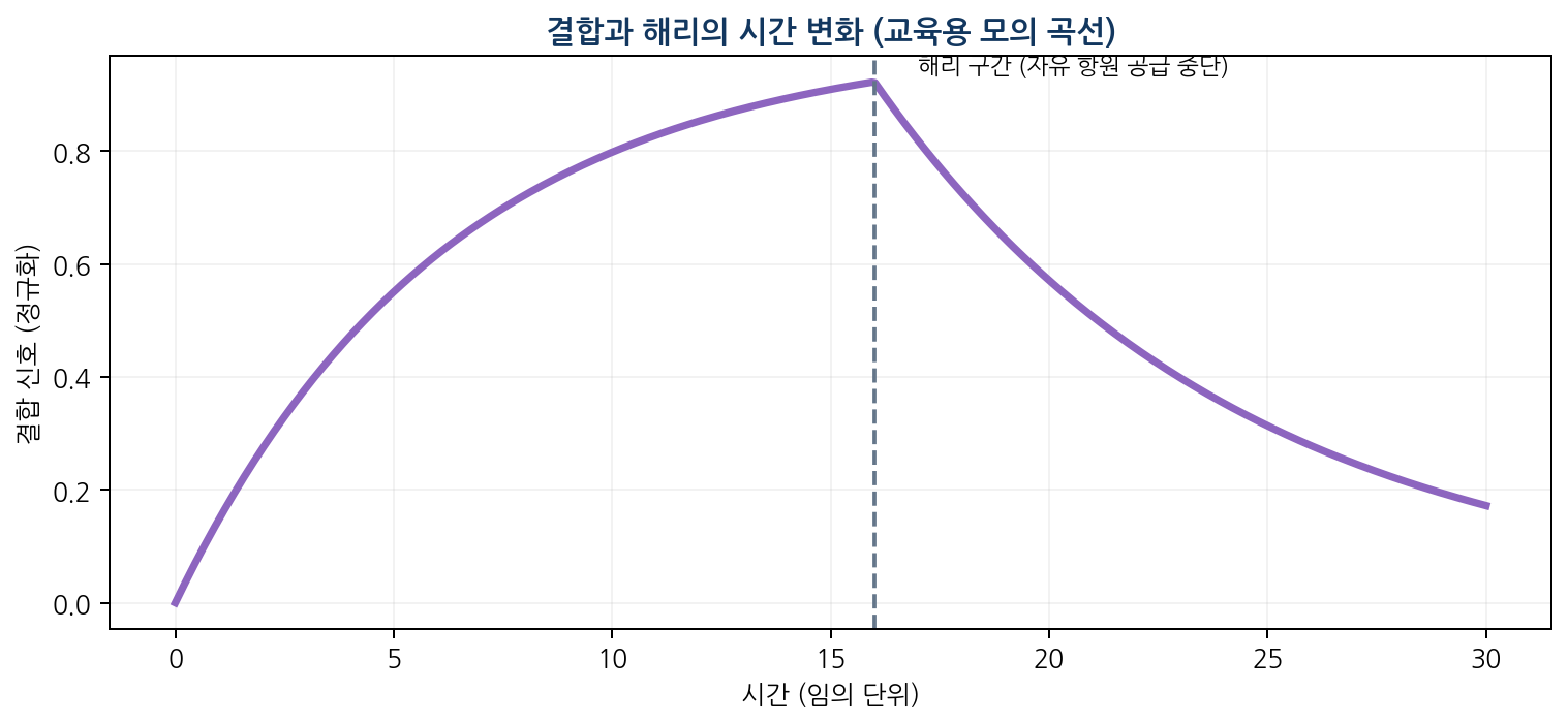
- kon has units M⁻¹s⁻¹ and koff units s⁻¹; two antibodies with the same Kd can still associate and dissociate at different rates
- Example: kon 2.0×10⁵ and koff 2.0×10⁻⁴ give Kd 1.0×10⁻⁹ M, and t₁/₂ = ln2/koff
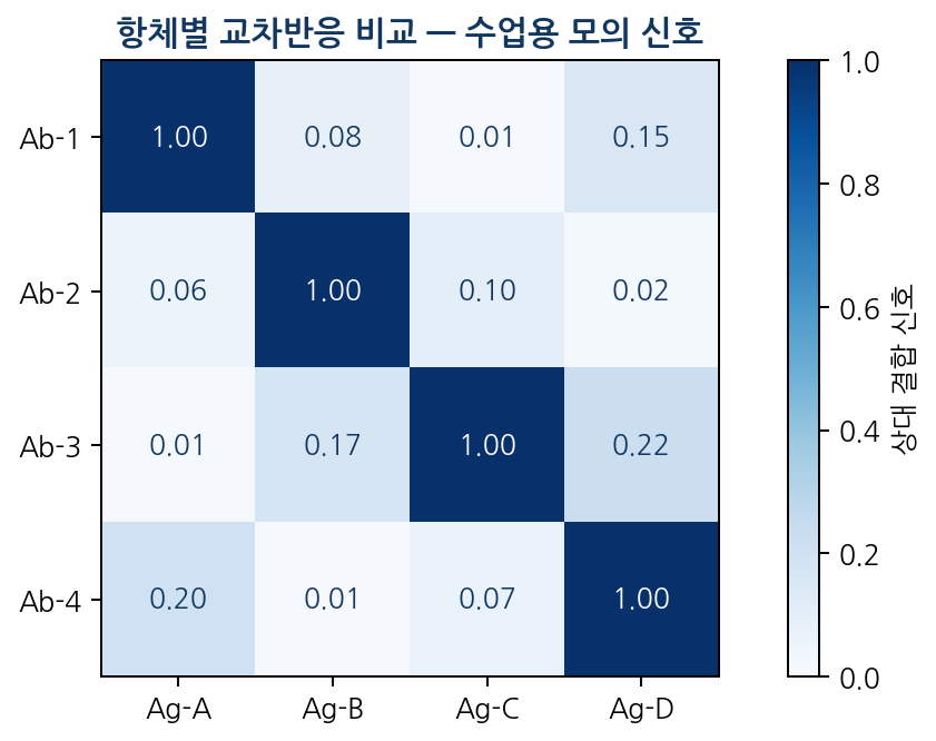
- Cross-reactivity is partial binding to similar epitopes; high concentrations of similar proteins, matrix interference and non-specific adsorption cause unexpected signal
- Specificity validation is essential when choosing a detection antibody
04
Antigen–antibody interactions underpin important assays
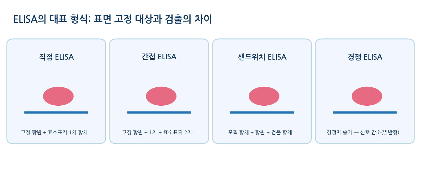
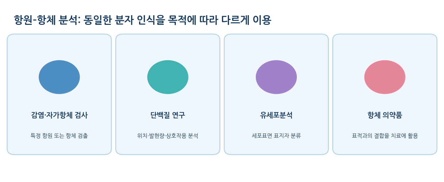
- ELISA reads antigen–antibody complex formation through an enzymatic colour reaction, in direct, indirect, sandwich or competitive format
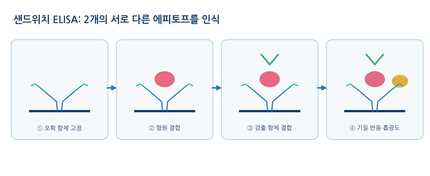
- A sandwich assay uses capture and detection antibodies that recognise different parts of the target
- In a competitive ELISA the signal falls as target concentration rises
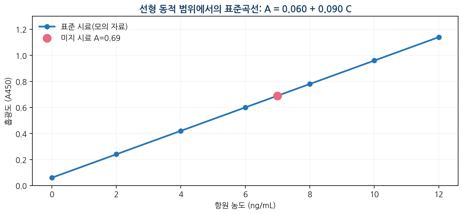
- Blocking and washing at each step remove non-specific binding and excess reagent; stay within the validated range of the standard curve
- Example: A450 0.690 with A = 0.060 + 0.090C gives 7.0 ng/mL, so a 5-fold dilution corresponds to 35 ng/mL in the original sample
- Lateral flow devices form test (T) and control (C) lines as labelled particles move through a porous membrane
- The control line mainly confirms sample migration and device function, and reading rules differ between products
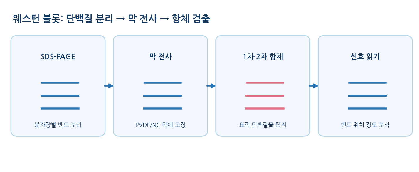
- Western blot separates proteins by electrophoresis, transfers them to a membrane and detects with primary and labelled secondary antibodies; position gives approximate mass and intensity a relative amount
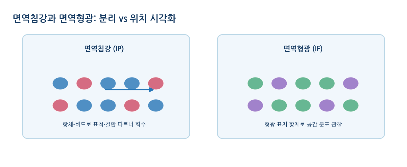
- Immunoprecipitation recovers a target with binding partners, immunofluorescence shows location and flow cytometry compares markers across many cells
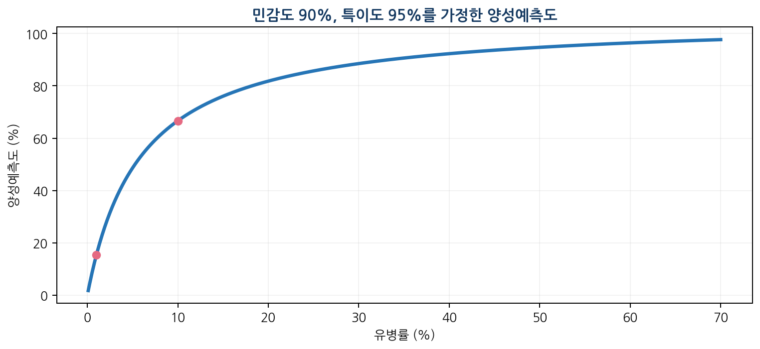
- Diagnostic sensitivity = TP/(TP+FN), specificity = TN/(TN+FP) and positive predictive value = TP/(TP+FP)
- Example: prevalence 1%, sensitivity 90%, specificity 95% gives 90 true and 495 false positives per 10,000, so PPV ≈ 15.4%
- Reliability requires positive and negative controls, blanks, standards, replicates and a defined measuring range
- Distinguish analytical specificity from clinical specificity, and limit of detection (LOD) from limit of quantification (LOQ)
- A monoclonal antibody comes from one clone with a single specificity: consistent, but vulnerable to epitope change or masking
- A polyclonal antibody is a mixture from several clones: robust signal from multiple epitopes, but batch variation and cross-reactivity need care
References
| Component | Representative cells and molecules | Core role |
|---|---|---|
| Innate immunity | Neutrophils, macrophages, dendritic cells, NK cells, complement | Early defence, antigen presentation, pathogen clearance |
| Humoral adaptive immunity | B cells, plasma cells, antibodies | Neutralisation, tagging and clearance of extracellular targets |
| Cell-mediated adaptive immunity | CD4⁺ helper T cells, CD8⁺ cytotoxic T cells | Regulation of the response; killing of infected cells |
| Immunological memory | Memory B cells, memory T cells | Rapid and efficient response on re-exposure |
| Isotype | Typical form | Main biological role |
|---|---|---|
| IgG | Mostly monomer (two binding sites) | Neutralisation and opsonisation in blood and tissue; some complement activation; placental transfer |
| IgM | Secreted form mostly pentameric | Early response; high avidity; complement activation |
| IgA | Secreted form mostly dimeric | Mucosal and secretion defence in gut and airway |
| IgE | Monomer | Mast cell and basophil Fc receptors; allergy and parasite defence |
| IgD | Mostly monomer | Mainly the antigen receptor of mature naive B cells |

| Method | Main target observed | Strength | Typical limitation |
|---|---|---|---|
| Sandwich ELISA | Concentration of the target antigen | Quantitative; many samples | Two antibodies must bind different epitopes at once |
| Indirect ELISA | Specific antibody in the sample | Antibody-response testing | Cross-reactivity; timing of sampling |
| Rapid antigen test | Presence of target (partly semi-quantitative) | Fast point-of-care reading | Limit of detection; sampling; product-specific reading |
| Western blot | Specific protein band | Detection with mass information | Off-target bands; non-linear quantification |
| Immunofluorescence | Spatial distribution of the target | Visualises cellular location | Depends on antibody, fixation and permeabilisation |