01
Actin and myosin are the main motor proteins of muscle
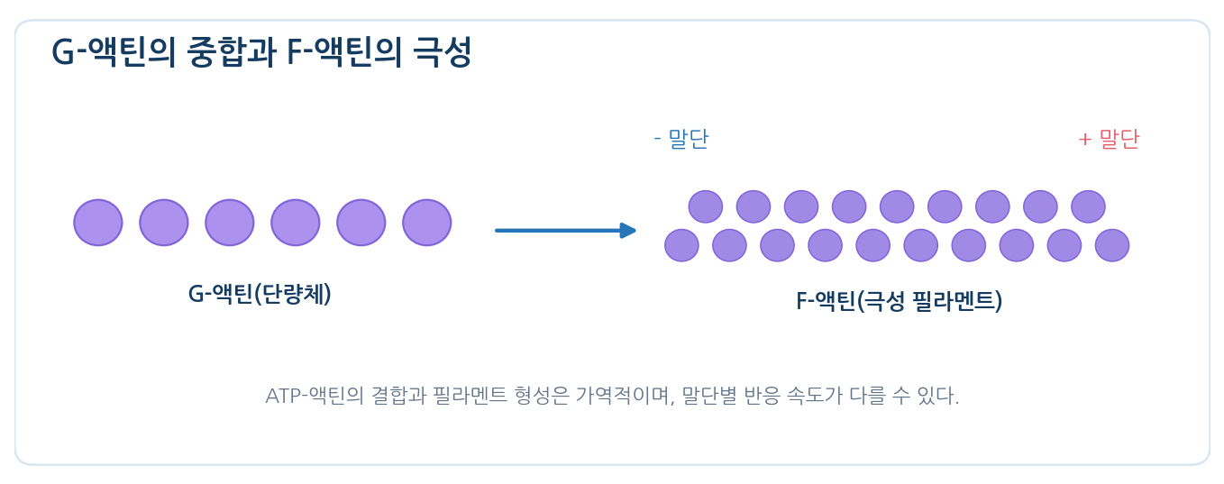
- G-actin is a globular monomer of about 42 kDa; polymerisation gives F-actin
- F-actin is a two-stranded helical microfilament about 7 nm thick, with structurally and kinetically distinct (+) and (−) ends
- G-actin binds ATP or ADP, and polymerisation and depolymerisation underlie cytoskeletal dynamics
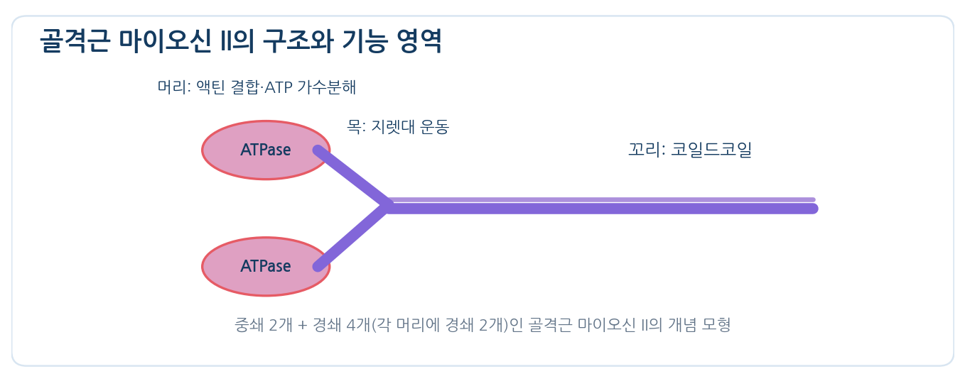
- Myosin II has two heavy chains and four light chains; the skeletal isoform is roughly 500 kDa
- The head carries the actin-binding site and the ATPase site; the neck acts as a lever that amplifies small rotations
- The two coiled tails build the backbone of the thick filament
02
Other proteins keep the filaments in ordered arrays
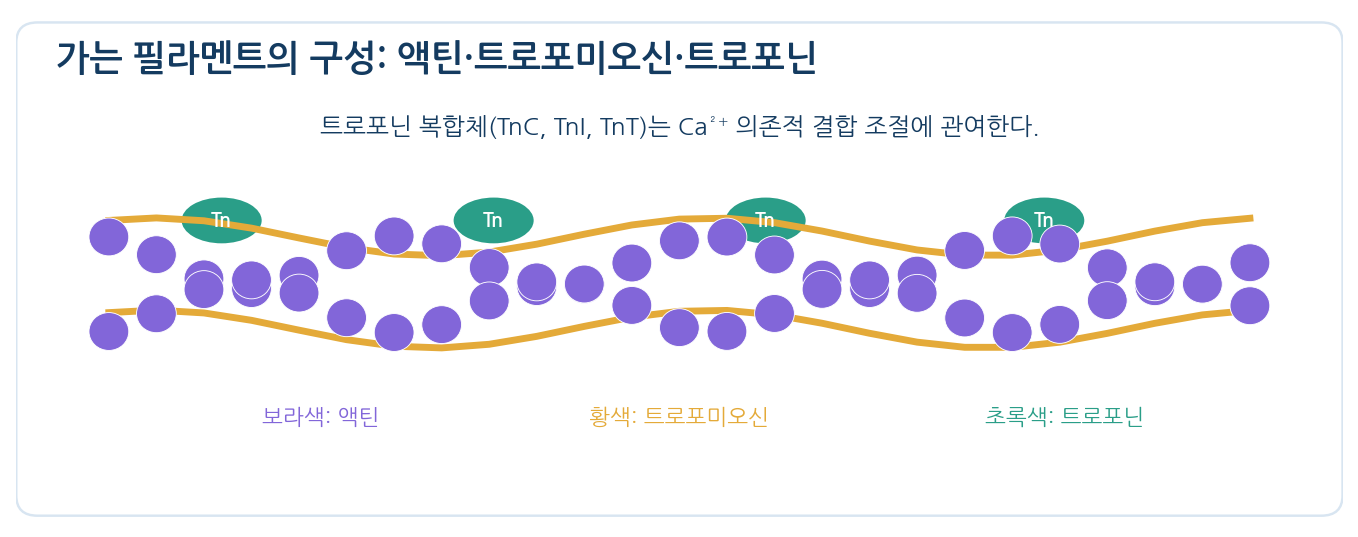
- The thin filament of striated muscle carries tropomyosin and troponin in addition to actin
- Tropomyosin lies along actin and restricts access to myosin-binding sites
- Troponin has three parts: TnC binds Ca²⁺, TnI inhibits and TnT binds tropomyosin
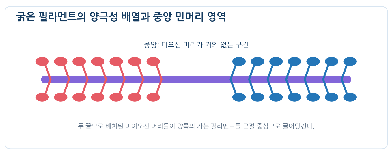
- Thick filaments bundle tails so heads point in both directions, leaving a bare zone at the centre
- Opposing heads pull thin filaments from both Z discs toward the M line
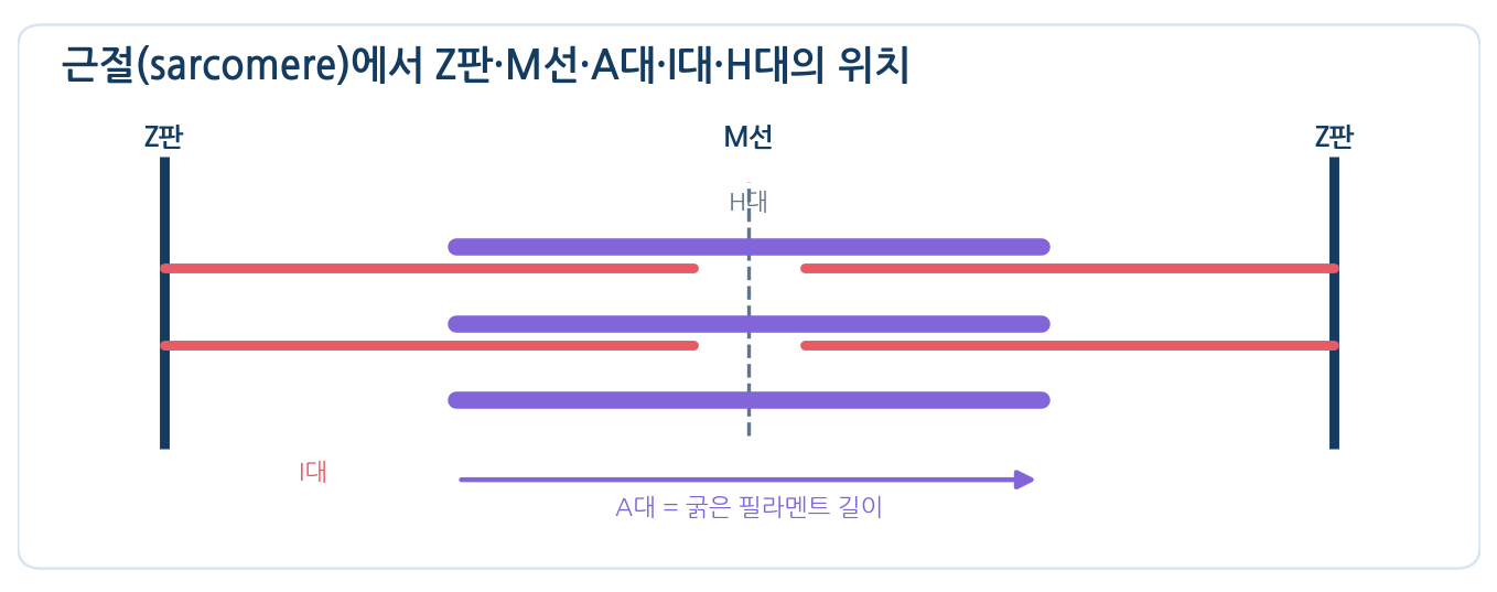
- The sarcomere is the unit between adjacent Z discs; the A band is the thick-filament region and keeps its length
- The I band contains only thin filaments, the H zone only thick filaments, and the M line aligns and links thick filaments
03
Relative sliding of myosin and actin shortens the muscle
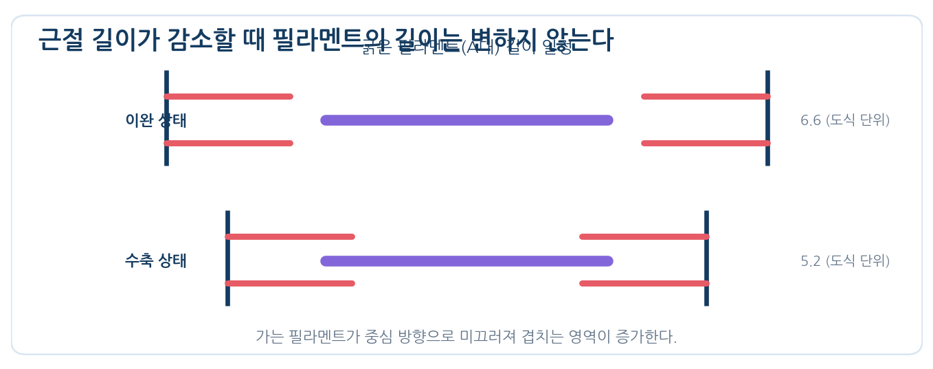
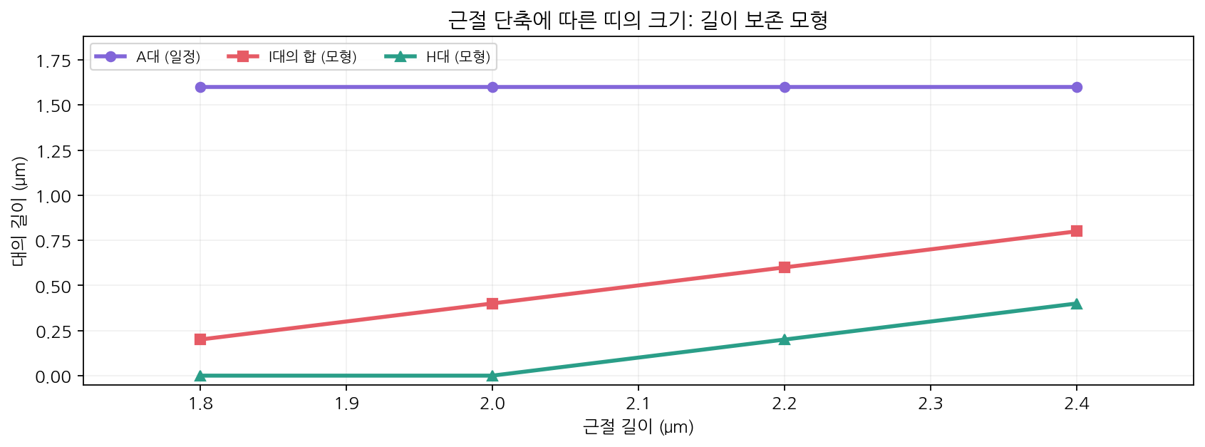
- On contraction the Z discs approach, the A band stays nearly constant and the I band and H zone shorten
- The band change reflects increased overlap, not shortening of the filaments themselves
- A cross-bridge is a myosin head bound to actin
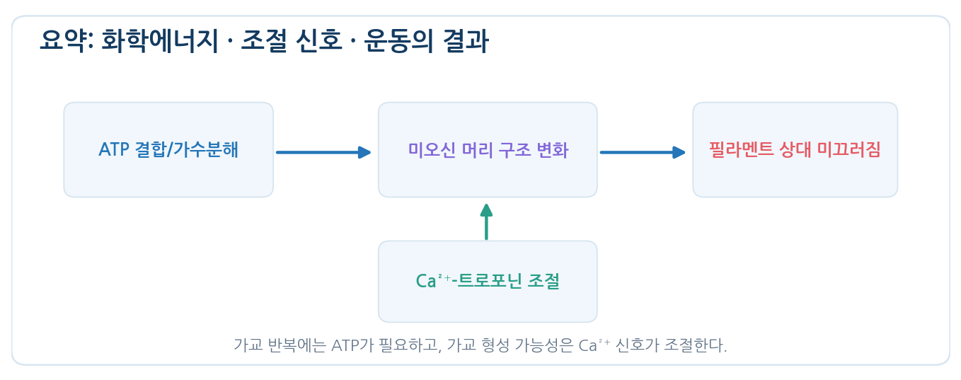
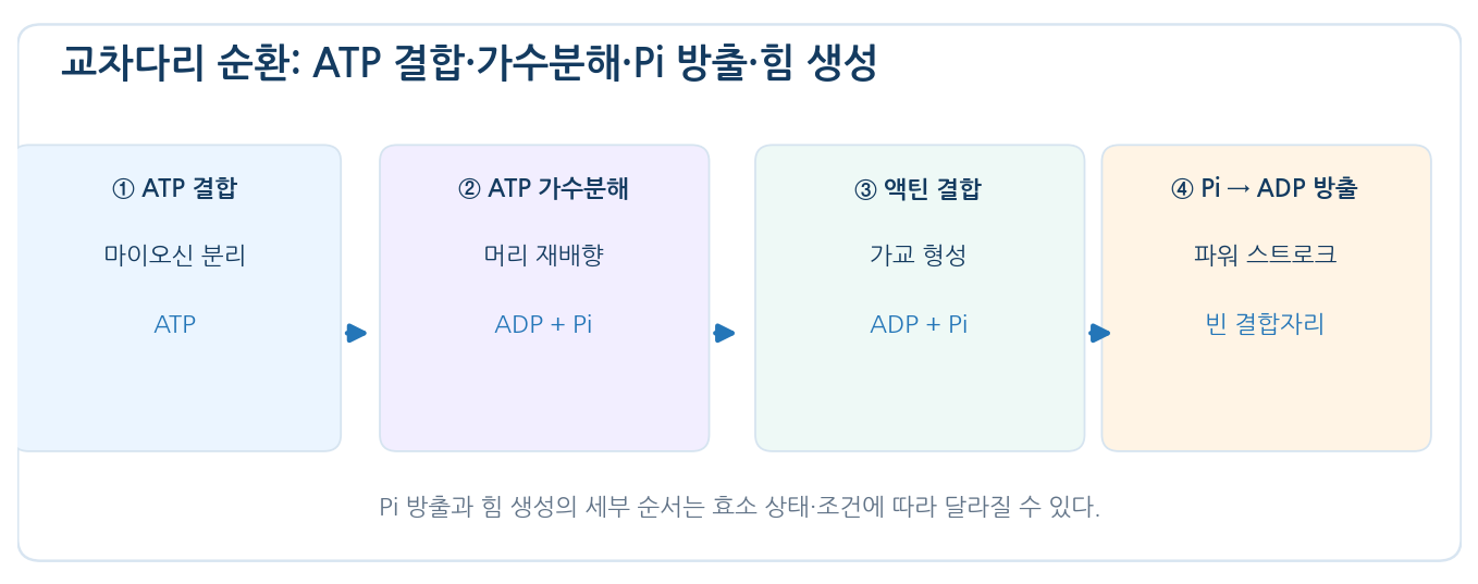
- Cross-bridge cycle: ATP binding, detachment, re-cocking after hydrolysis, actin binding, Pi release and force, then ADP release
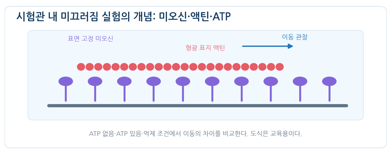
- Repeated cycles produce relative filament motion; without ATP the heads cannot detach from the strong-binding state
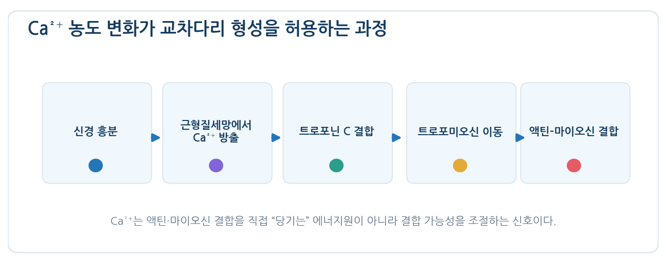
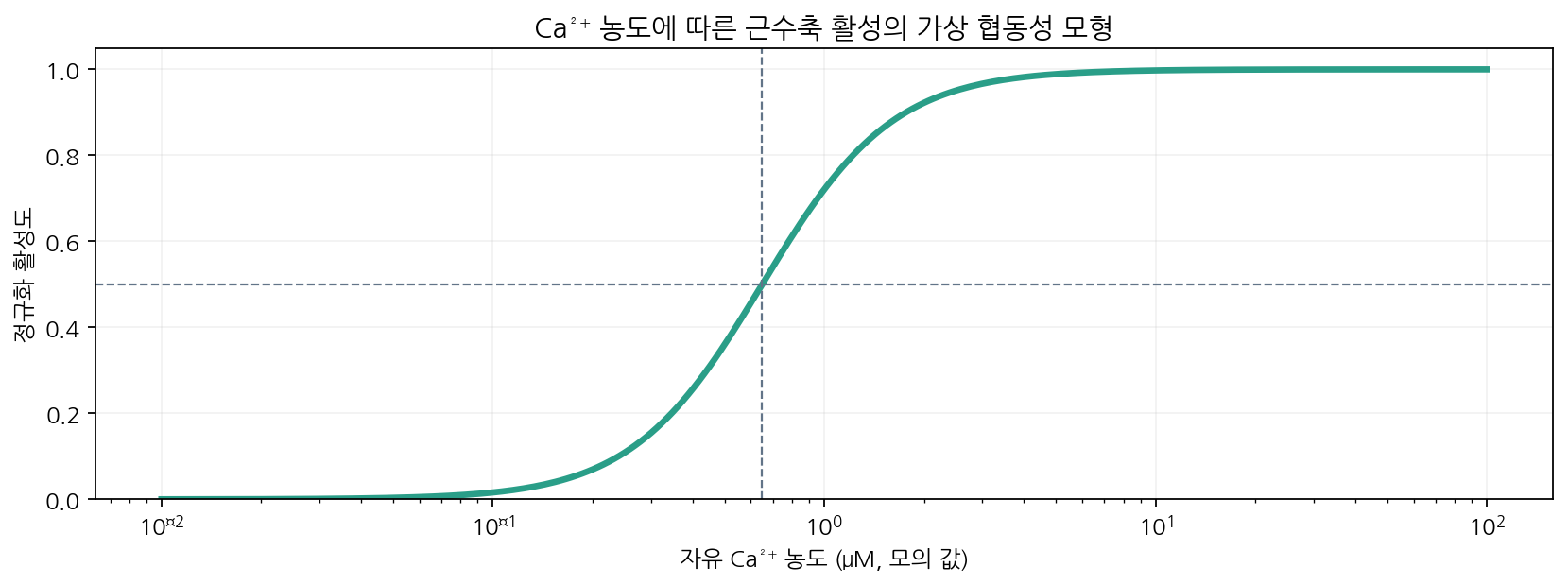
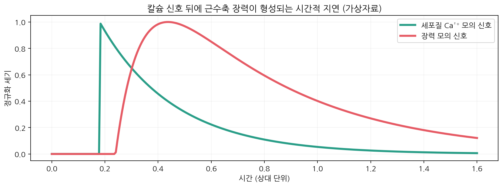
- Nerve stimulation releases Ca²⁺ from the sarcoplasmic reticulum, TnC binds it, tropomyosin moves and sites are exposed
- When stimulation ends, SERCA Ca²⁺-ATPase pumps Ca²⁺ back at the cost of ATP and relaxation follows
- Length–tension relation: over-stretching reduces available bridges and over-shortening causes interference, both lowering active tension
- Passive tension from titin and connective tissue is considered separately
04
Chemical energy is converted into mechanical work
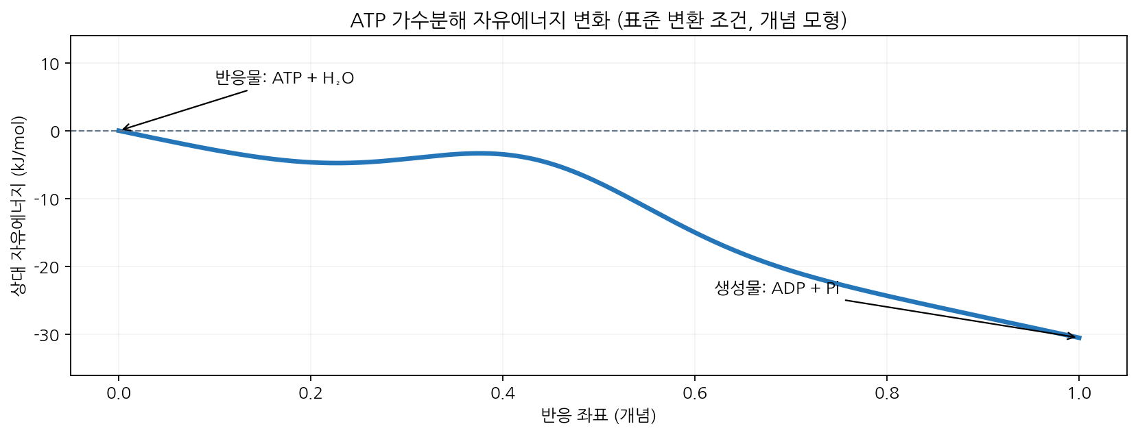
- The standard free-energy change of ATP + H2O → ADP + Pi is about −30.5 kJ/mol (25 °C, pH 7)
- The cellular value is usually more negative, and ΔG = ΔG°′ + RT ln([ADP][Pi]/[ATP])
- A negative ΔG means the overall reaction is favourable; bond cleavage does not create energy
- Myosin couples ATP binding, hydrolysis and product release to structural change
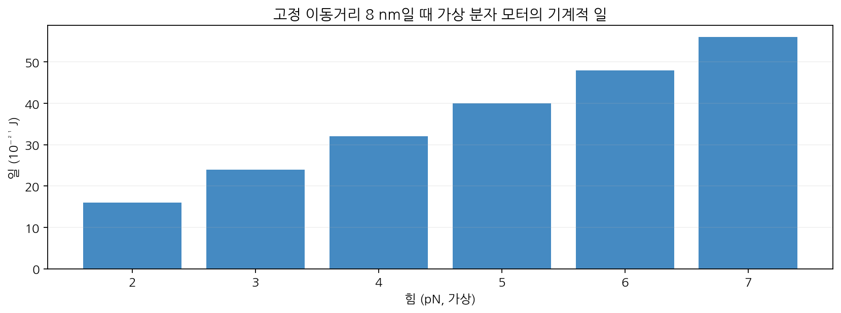
- Work W = Fd, with forces in pN and distances in nm, so energies are of order 10⁻²¹ J
- Efficiency η = W/(|ΔG_ATP|/N_A) × 100 depends on conditions and on how work is defined
- Example: 5 pN over 8 nm gives 4.0×10⁻²⁰ J, about 48% efficiency if ΔG is taken as −50 kJ/mol
- Rigor mortis reflects ATP depletion together with loss of ion homeostasis, not simply stronger contraction
05
The principle of the molecular motor extends beyond muscle
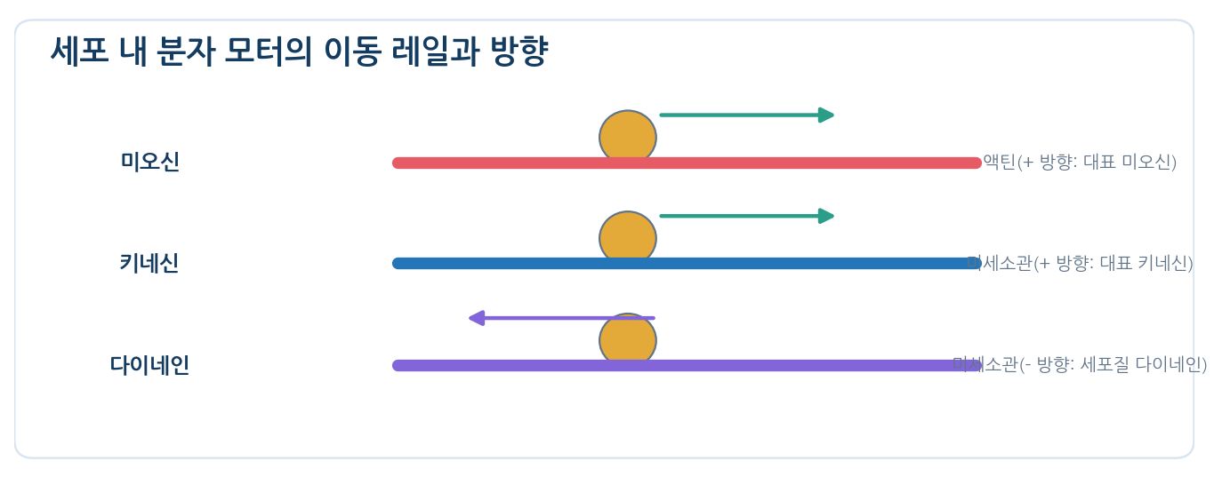
- Myosin V carries intracellular cargo along actin
- Kinesin and dynein mostly use microtubules as tracks
- Many conventional kinesins move toward the (+) end, while cytoplasmic dynein moves mainly toward (−)
- Not every kinesin or myosin moves in the same direction or walks the same way; exceptional isoforms exist
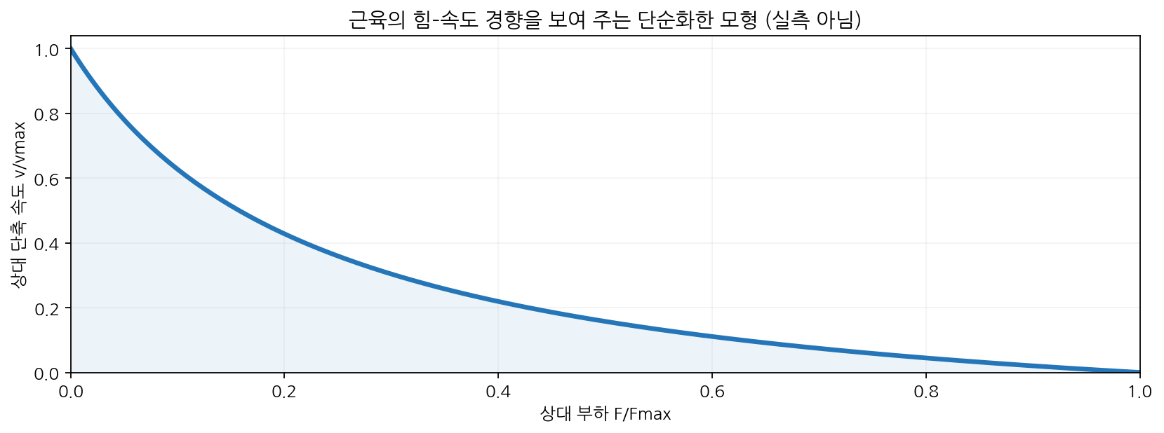
- Contraction velocity falls as load rises, and mechanical work can be computed from ATP use and distance moved
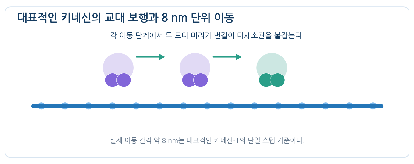
- The shared principle is a directional motor cycle that couples ATP turnover to binding, detachment and re-cocking
References
| Feature | Thin filament | Thick filament |
|---|---|---|
| Core protein | F-actin | Myosin II |
| Typical diameter | About 7 nm | About 15 nm |
| Characteristic binding and function | Myosin-binding surface; structural polarity | ATPase heads; force generation |
| Structural arrangement | Anchored at the Z disc, extending toward the centre | Bipolar array around the M line |
| Accessory proteins | Troponin, tropomyosin and others | Titin, MyBP-C and others |
| Accessory protein | Location or function | Points to remember |
|---|---|---|
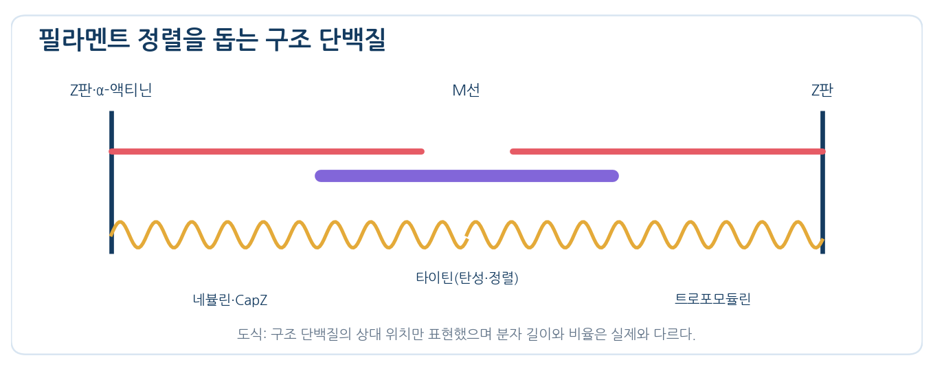
| α-actinin / CapZ | Aligns and anchors actin at the Z disc / caps the (+) end | The Z disc is the anchor of the thin filament |
| Tropomodulin | Caps the (−) end of the thin filament | Controls length and stability |
| Nebulin | Binds along the thin filament | Contributes to length control (varies with muscle) |
| Titin | Giant elastic protein linking the Z disc toward the M line | Thick-filament position and passive elasticity |
| MyBP-C | Binds the thick filament | Modulates myosin activity and packing |
| ATP / product state | Myosin–actin relationship | Key interpretation for teaching |
|---|---|---|
| ATP bound | Actin affinity falls and the head detaches | ATP binding is needed for detachment |
| ADP and Pi bound | The head re-cocks into a position favourable for the next binding | Couples hydrolysis to a structural change |
| Pi release and strong binding | Linked to the force-generating step | Main transition of the power stroke |
| After ADP release | The head can stay strongly bound | Without the next ATP the cycle stalls |
| Motor protein | Track | Typical movement | ATP and role |
|---|---|---|---|
| Muscle myosin II | Actin | Many heads generate force collectively | Sarcomere contraction |
| Myosin V | Actin | Long walking strides carrying organelles | Intracellular cargo transport |
| Kinesin-1 | Microtubule | Mainly toward (+), about 8 nm per step | Vesicle and organelle transport |
| Cytoplasmic dynein | Microtubule | Mostly toward (−), irregular steps | Retrograde transport and more |