01
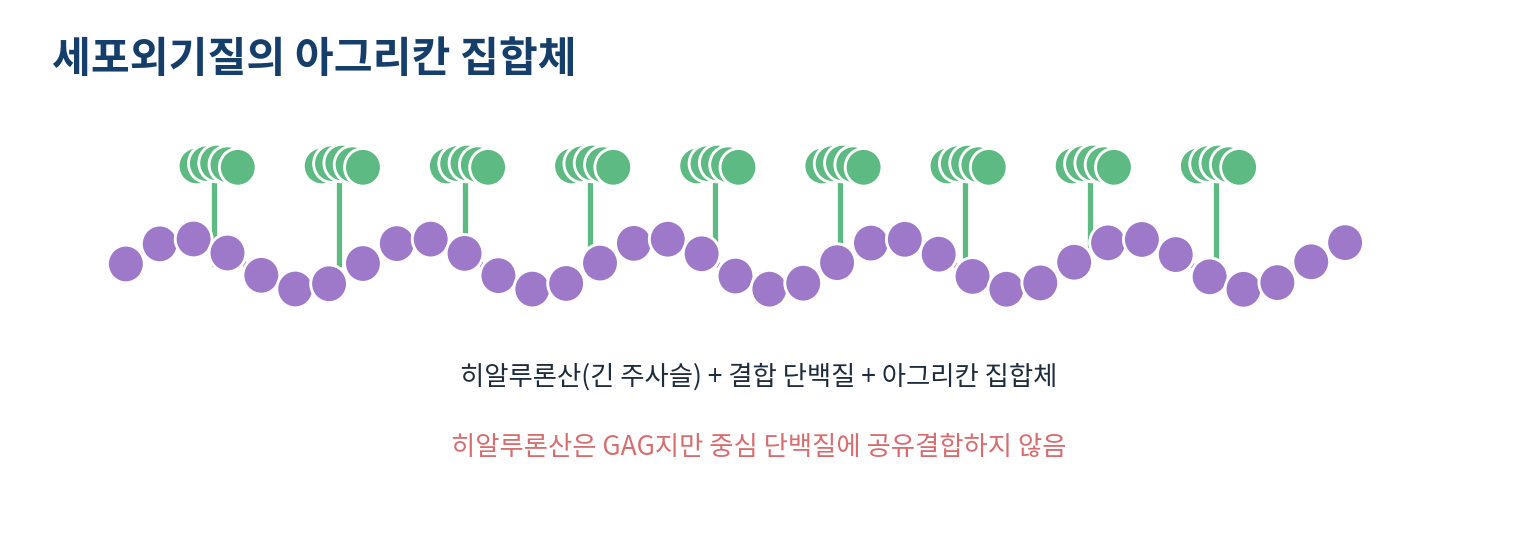
Proteoglycans carry GAG chains at the cell surface and in the matrix
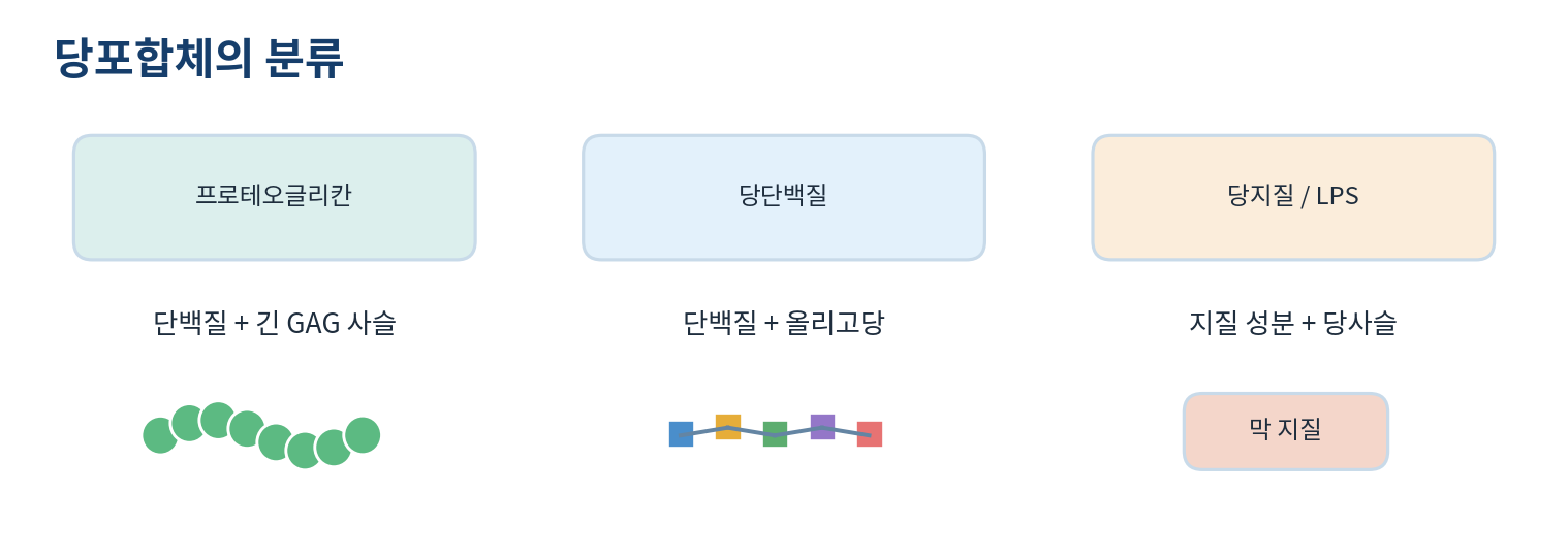
- Glycoconjugates are molecules in which glycans are joined to proteins or lipids
- Glycan structure is set by sugar type, linkage position, branching, terminal sugar and sulfation
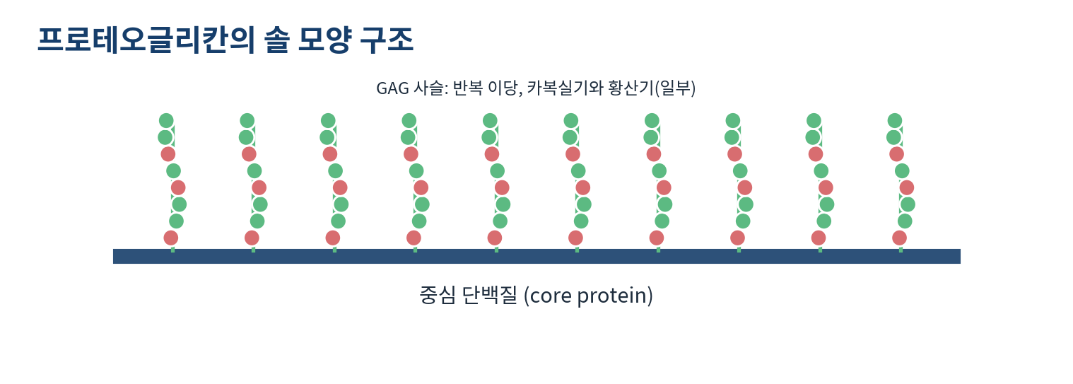
- A proteoglycan is a core protein carrying covalently attached GAG chains
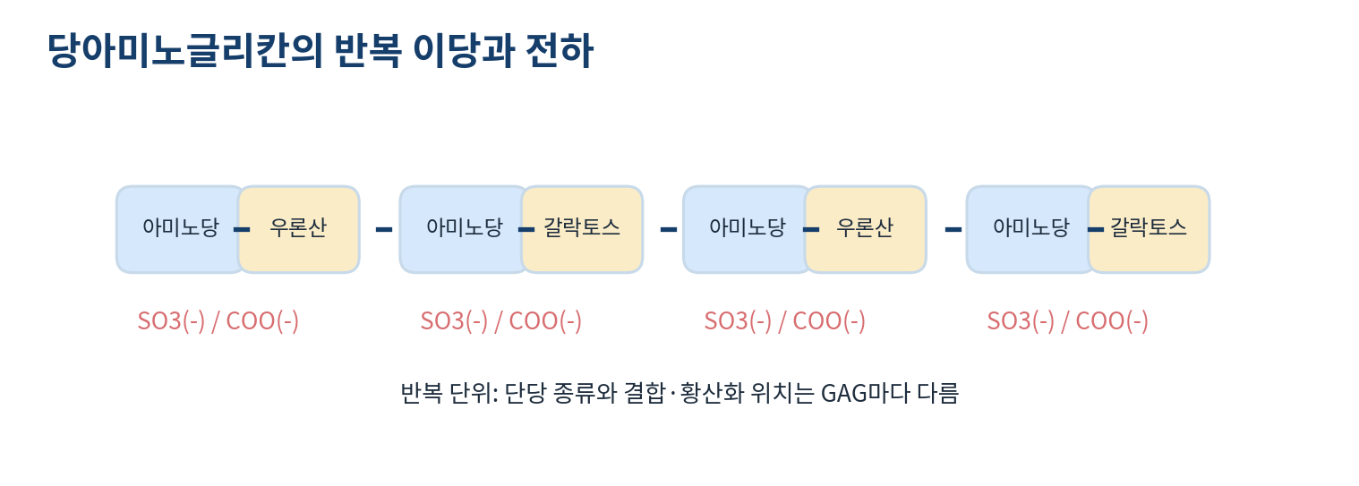
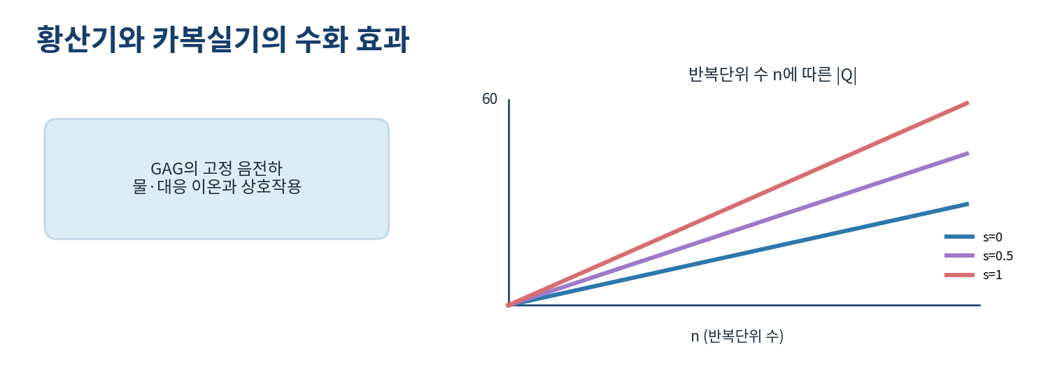
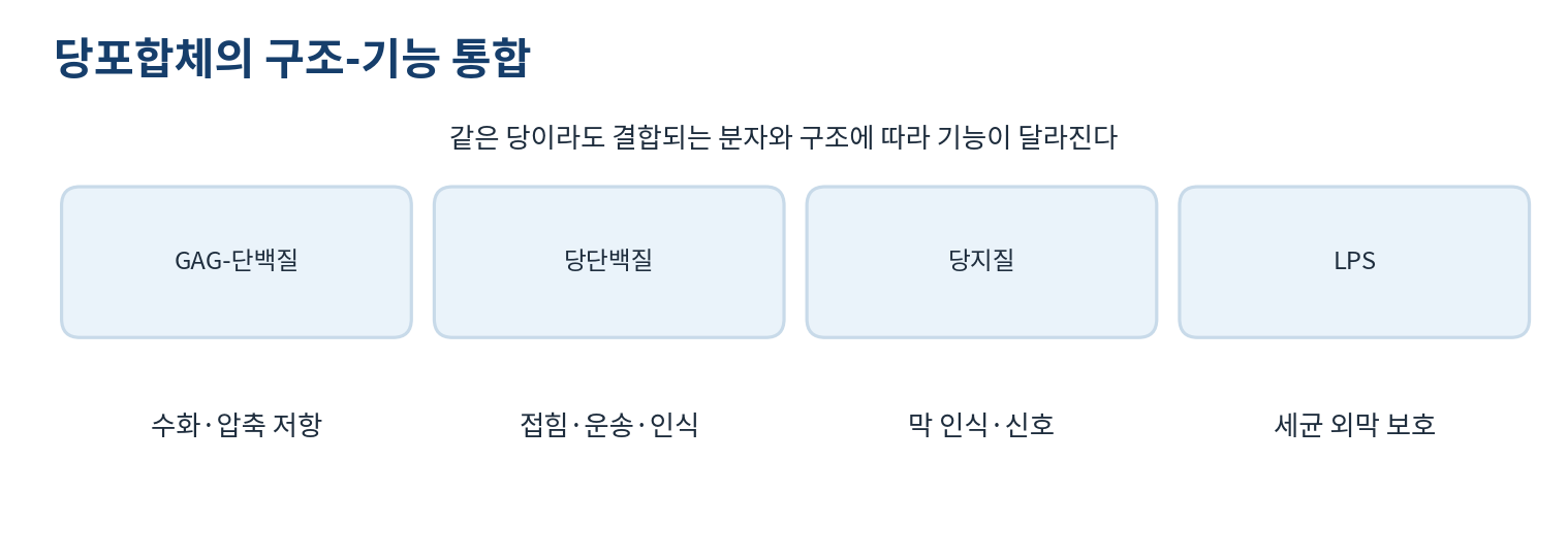
- GAGs are repeating disaccharides of an amino sugar with a uronic acid or galactose, and carboxylate and sulfate charges support hydration and compression resistance
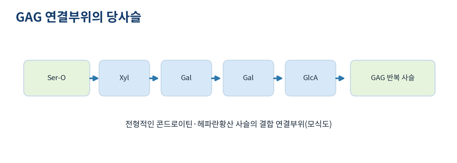
- Chondroitin and heparan sulfate attach through serine to xylose, two galactoses and glucuronic acid
- That linkage cannot be generalised to every GAG
- Aggrecan carries many chondroitin and keratan chains and binds hyaluronan non-covalently to form large aggregates
- Example: with 20 repeats each carrying one carboxyl and 0.8 sulfate the mean formal charge is about -20 x 1.8, that is -36
02
[Separate note 7-3] Disorders of sulfated GAG turnover
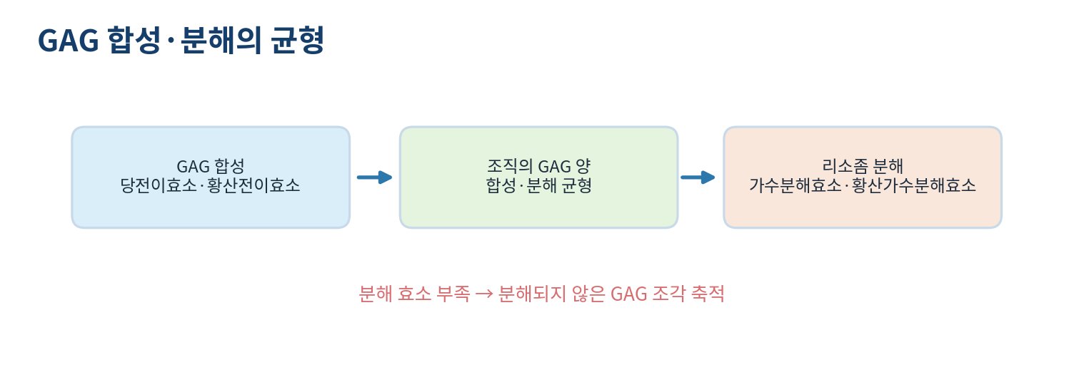
- GAG amount reflects a balance of biosynthesis, degradation and extracellular traffic
- Defects in glycosyltransferases or sulfotransferases change chain length or sulfation and affect tissue formation and signalling
- Deficiency of lysosomal hydrolases or sulfatases lets GAG fragments accumulate
- MPS I results from reduced alpha-L-iduronidase activity through IDUA variants, impairing breakdown of dermatan and heparan sulfate
- Undegraded GAG accumulates in lysosomes and affects many tissues, with highly variable clinical features
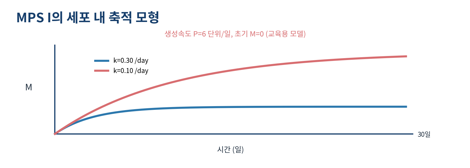
- The model dM/dt = P - kM gives M* = P/k, so P = 6 with k = 0.30 gives 20 units and k = 0.10 gives 60
- Those values are a teaching model, not measured patient data
03
Glycoproteins carry covalently linked oligosaccharides
- A glycoprotein carries glycans covalently attached to protein
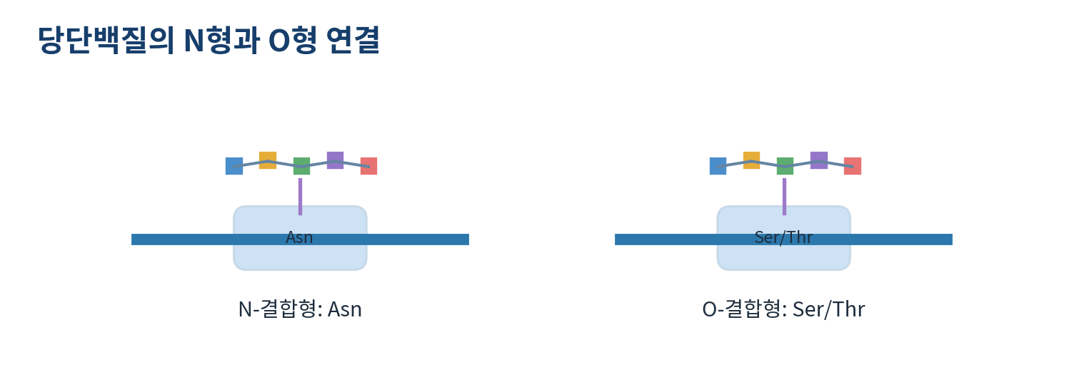
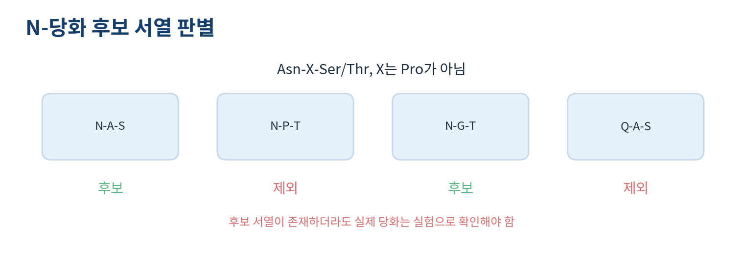
- N-linked glycosylation attaches GlcNAc to the amide nitrogen of asparagine, with the candidate sequon Asn-X-Ser/Thr where X is usually not proline
- A sequon alone does not guarantee glycosylation, since accessibility, structure and localisation also act
- O-linked mucin-type glycosylation starts with GalNAc on the hydroxyl of Ser or Thr
- Glycosylation is enzymatic, whereas glycation is the non-enzymatic reaction of a reducing sugar with protein
- Glycans support folding quality control, secretion, stability and recognition, and the many O-linked chains of mucin favour lubrication and protection
- Signals at 28.0 and 31.2 kDa in mass data suggest a difference of about 3.2 kDa in glycan content
04
Glycolipids and lipopolysaccharides anchor glycans in membranes
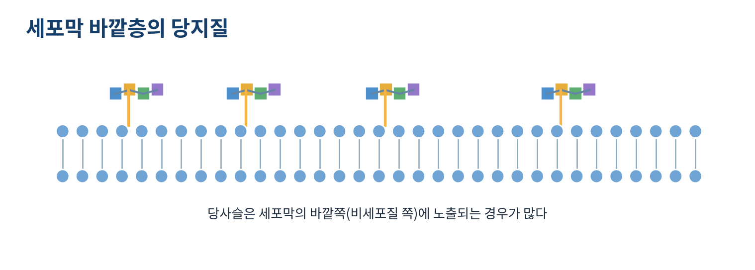
- A glycolipid carries glycan on a lipid, and in animal cells the glycosphingolipids built on ceramide are the main example
- Gangliosides are glycosphingolipids containing at least one sialic acid
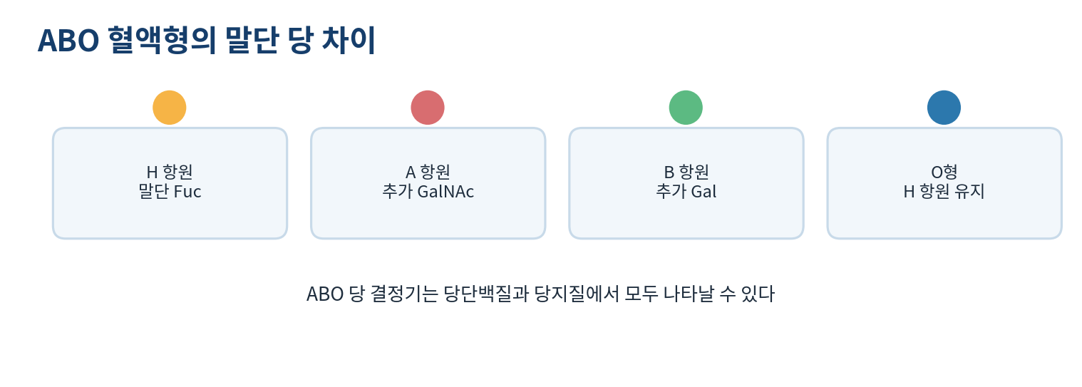
- ABO antigens differ in terminal sugar: adding GalNAc to the H antigen gives A and adding Gal gives B
- Type O usually retains the H antigen because the transferases that add the A or B terminal sugar are inactive
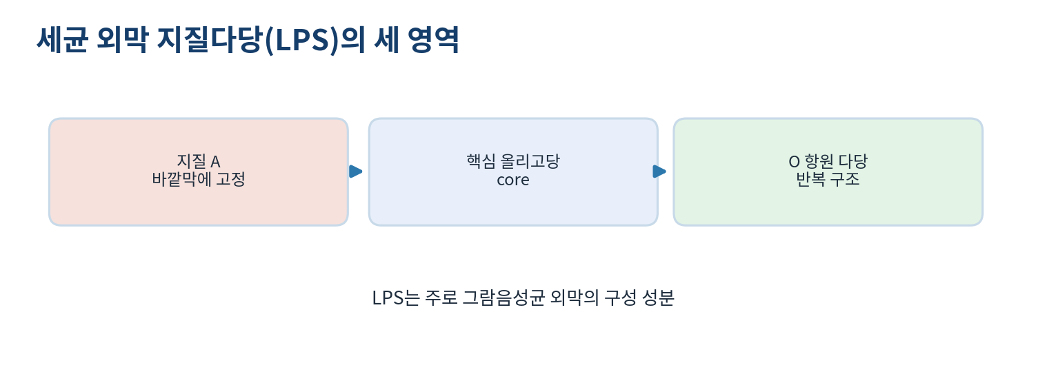
- Gram-negative lipopolysaccharide is lipid A for membrane anchoring plus a core oligosaccharide and the O antigen polysaccharide
- LPS and animal glycosphingolipids have different membrane anchors and cannot be placed in one family
05
Quantitative work and investigations
- The formal charge model for a GAG is Q = -n x (1 + s), where n is the number of repeats and s the mean sulfates per repeat
- With 20 repeats and 0.5 sulfate per repeat Q = -20 x 1.5 = -30
- The model ignores ionic strength, counterions and steric constraints, so it does not map one-to-one onto real charge, hydration or viscoelasticity
- In scanning for N-X-S/T the window with proline at X is excluded, and candidates are not guaranteed to be glycosylated
- The number of attached glycans changes mass and glycan mass fraction, and apparent electrophoretic mass can differ from mass spectrometry
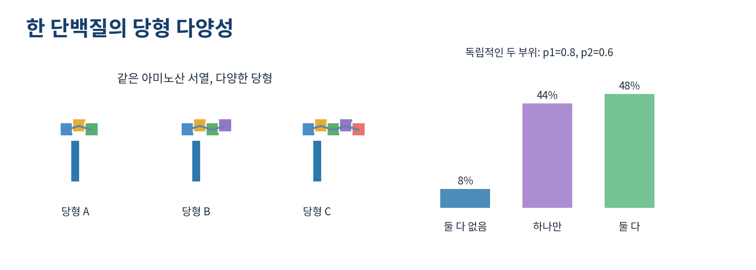
- One sequence gives several glycoforms through site occupancy, branching and cell-type-specific enzyme expression
References
| Family | Composition and linkage | Examples | Main function |
|---|---|---|---|
| Proteoglycan | Core protein with long GAG chains | Aggrecan, syndecan | Hydration, compression resistance, signalling |
| Glycoprotein | Protein with N- or O-linked oligosaccharides | Mucin, antibodies, membrane receptors | Recognition, folding, protein trafficking |
| Glycolipid | Lipid anchor with outward glycan | Glucosylceramide, GM1 | Membrane recognition, signalling, membrane properties |
| Lipopolysaccharide | Lipid A with core sugar and O antigen | Gram-negative outer membrane | Barrier function and immune recognition |
| Term | Normal model | Reduced degradation | Interpretation |
|---|---|---|---|
| Production rate P | 6 units per day | 6 units per day | Unchanged |
| Degradation constant k | 0.30 per day | 0.10 per day | Falls to one third |
| Steady state M* | 20 units | 60 units | Threefold rise |
| Feature | N-linked | O-linked (mucin type) |
|---|---|---|
| Linked residue | Asparagine (Asn) | Serine or threonine |
| First sugar | GlcNAc | GalNAc |
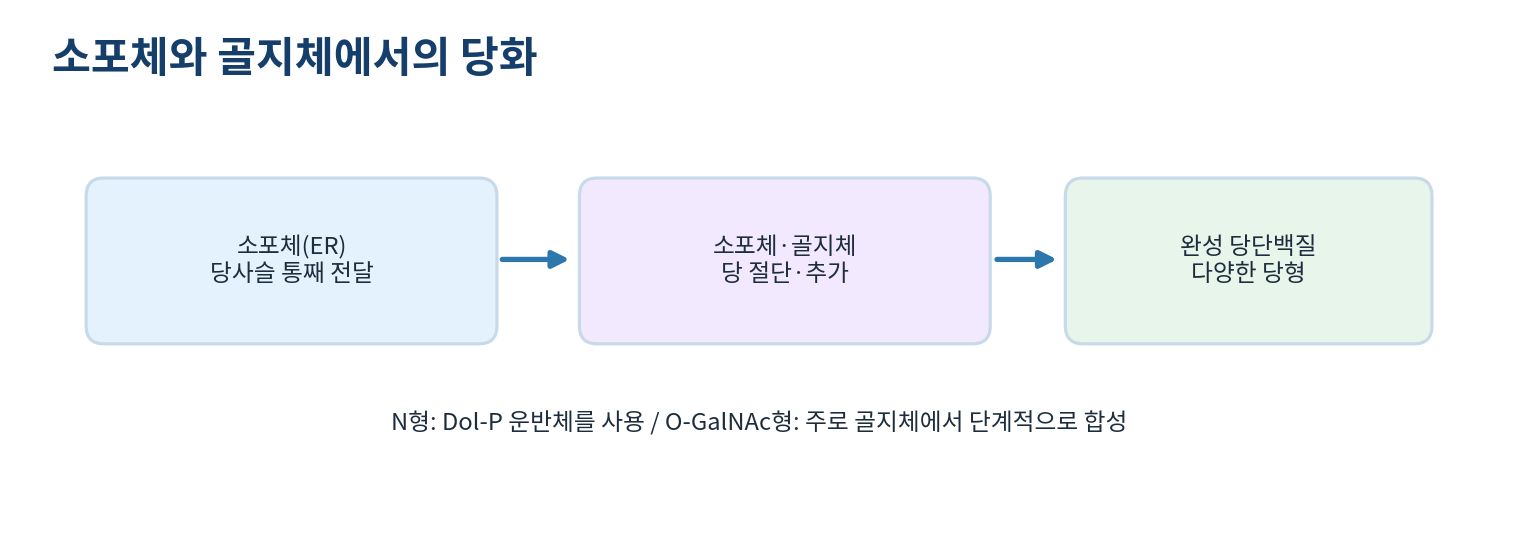
| Initial synthesis | Assembled on a lipid carrier in the ER then transferred | Usually added stepwise in the Golgi |
| Typical function | Folding quality control, secretion, recognition | Mucus formation, lubrication, surface protection |
| Caution | Asn-X-Ser/Thr is only a candidate and is not always glycosylated | Not every O-linked glycan is mucin type |
| Feature | Animal glycosphingolipid | Bacterial lipopolysaccharide |
|---|---|---|
| Membrane anchor | Ceramide | Lipid A |
| Typical location | Non-cytosolic leaflet of animal membranes | Outer leaflet of the Gram-negative outer membrane |
| Glycan structure | Various oligosaccharides on ceramide | Core sugar with repeating O antigen |
| Biological role | Cell recognition, signalling, membrane organisation | Barrier function, surface diversity, immune recognition |