01
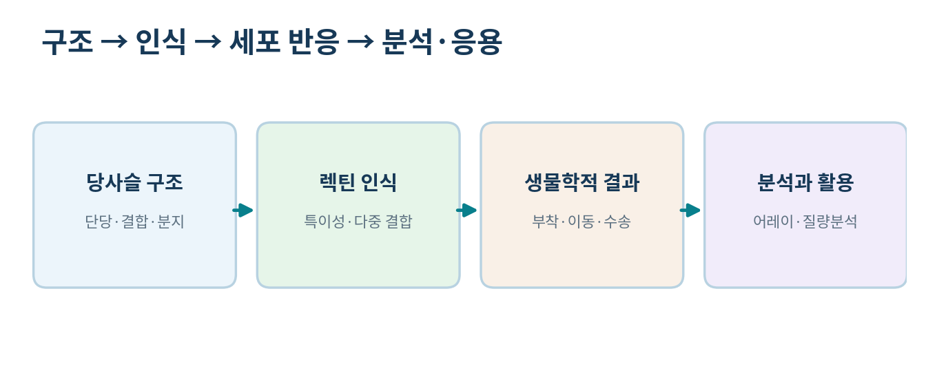
Glycans carry information through structural diversity
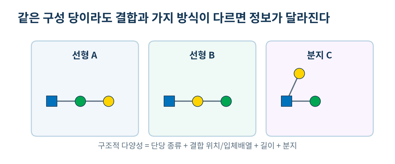
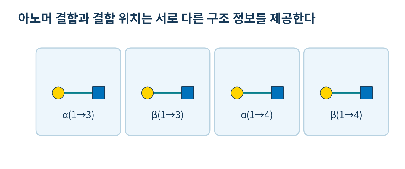
- Glycans differ by sugar type, anomeric configuration, linkage position, chain length, branching and acetylation or sulfation
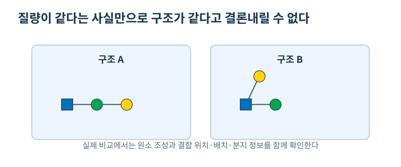
- Glycans of identical formula can interact differently with other molecules and therefore carry information
- Complex glycans are built in the ER and Golgi by enzymes that transfer sugars from nucleotide donors or trim existing chains
- The final structure depends on cell type, the distribution of glycosyltransferases and glycosidases, donor supply and trafficking
- The simple idea that one gene determines one glycan therefore fails
- The phrase sugar code is a metaphor for the whole recognisable structure, not a translation rule like the base sequence
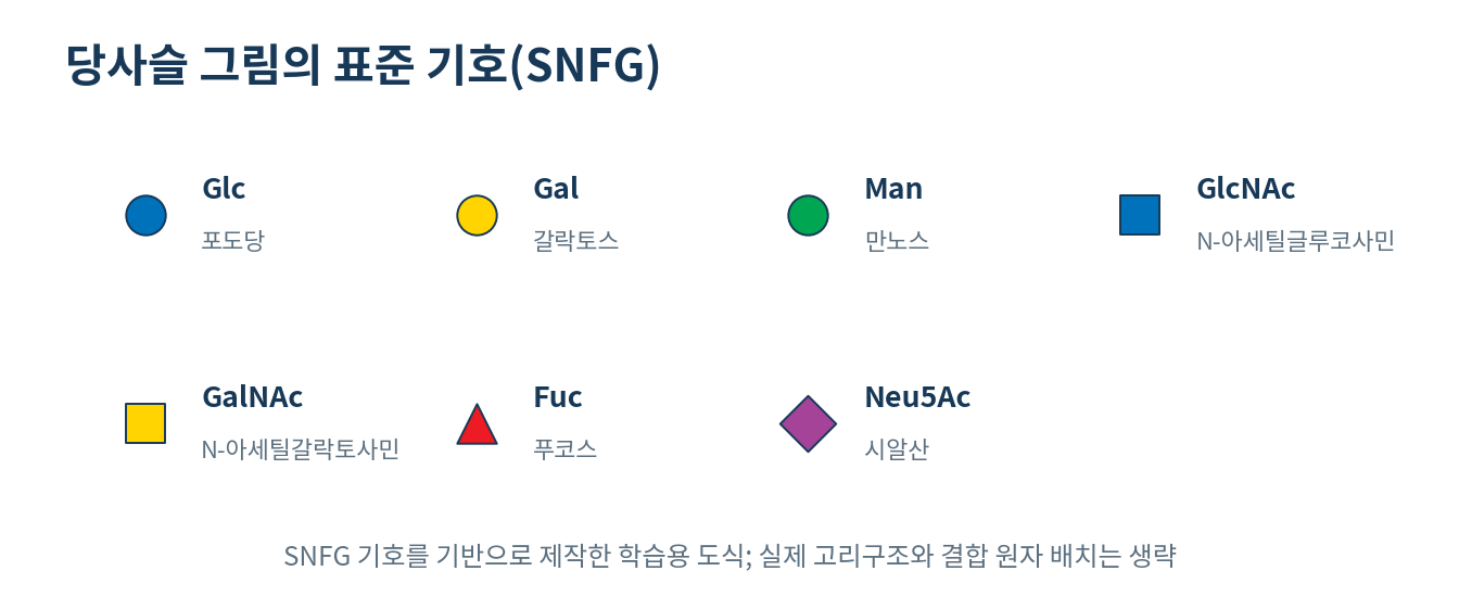
- SNFG assigns each monosaccharide a shape and colour so that complex structures can be drawn compactly, with blue circles for Glc, yellow for Gal and green for Man
02
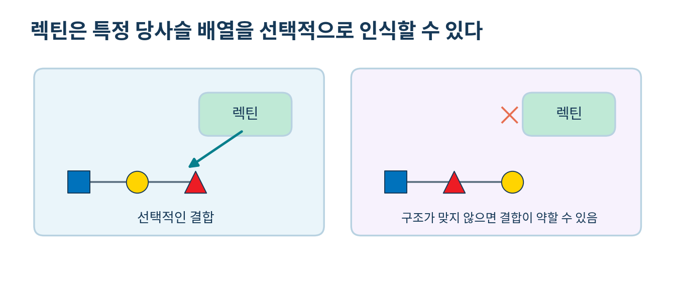
Lectins recognise glycan structure selectively
- Lectins are a large class of proteins that recognise and bind glycans, while glycan-binding protein is the broader term covering lectins, antibodies and microbial adhesins
- Binding rests on hydrogen bonds, dispersion forces, electrostatics, hydrophobic contacts and shape complementarity, and is mostly non-covalent
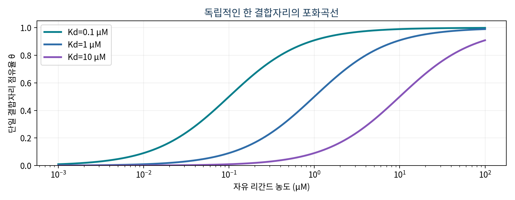
- For one site at equilibrium P + L to PL, Kd = [P][L]/[PL] and occupancy theta = [L]/(Kd + [L])
- A smaller Kd gives higher occupancy at the same concentration, and [L] equals Kd when theta is 0.5
- Example: Kd 0.50 micromolar with 2.0 micromolar ligand gives theta = 0.80
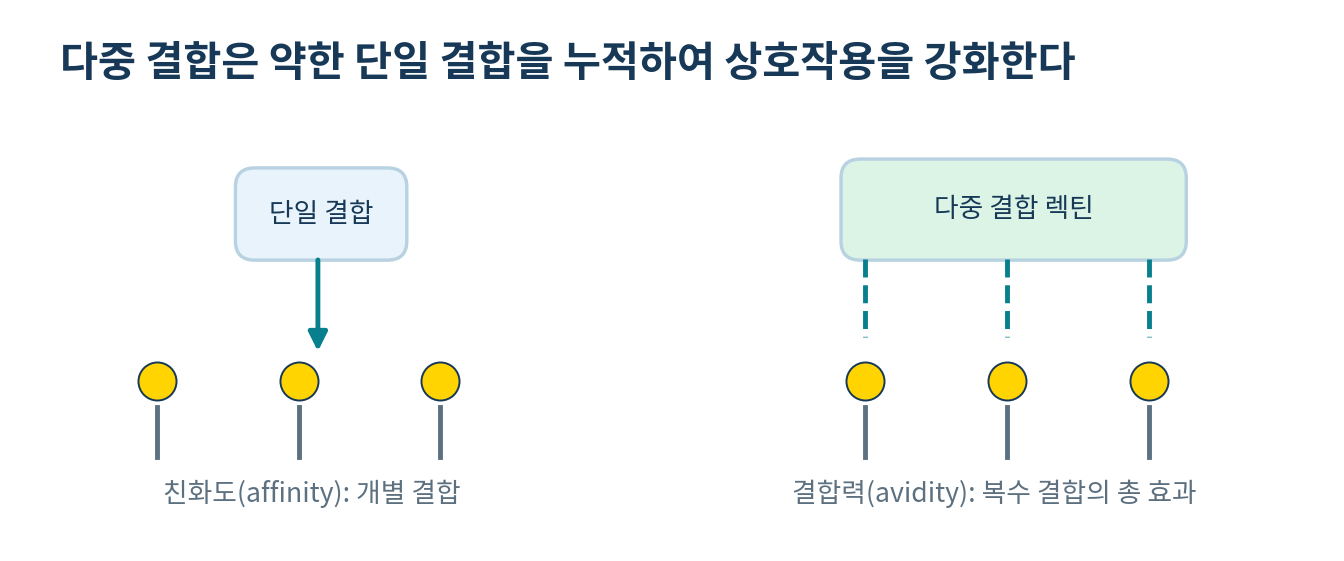
- Affinity at a single site and avidity across several sites are separate concepts
- When several sites engage at once, a broken contact can re-form nearby, which raises total avidity
03
Glycan recognition underlies adhesion, trafficking and immunity
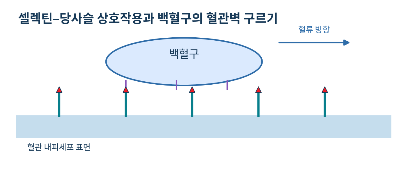
- Leukocytes first roll along the endothelium through weak and repeated binding before reaching an inflammatory site
- Selectins and glycan determinants on glycoprotein ligands act in this step, while integrins mediate firm adhesion
- Sialyl Lewis x contains sialic acid and fucose, yet functional selectin binding also depends on how the glycan is presented and on neighbouring sulfated residues
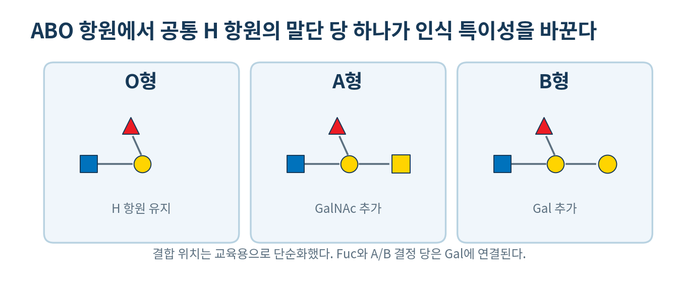
- ABO antigens arise by adding GalNAc to the H antigen for A and Gal for B, while type O lacks the transferase and keeps H
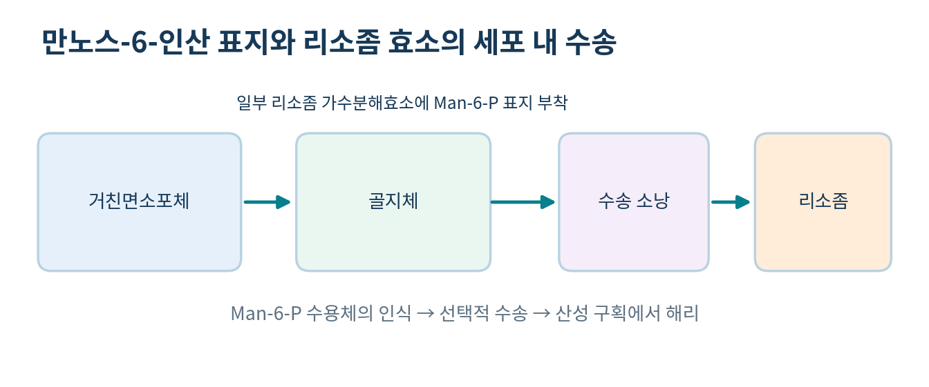
- Mannose 6-phosphate marks many lysosomal hydrolases for sorting from Golgi to endosome, though not every lysosomal protein uses only this route
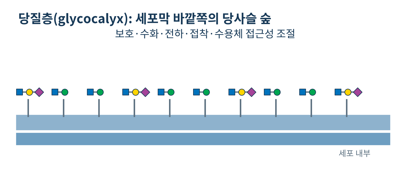
- The glycocalyx contributes hydration, surface charge, physical contact, receptor accessibility and cell interaction, with composition and thickness varying by cell
04
Microbes can recognise host surface glycans
- Viruses and bacteria can use particular host glycans as attachment targets
- Attachment influences the first stage of infection, but protein receptors, host defence and tissue environment also determine whether infection establishes
- Binding to a glycan does not by itself establish infectivity or virulence
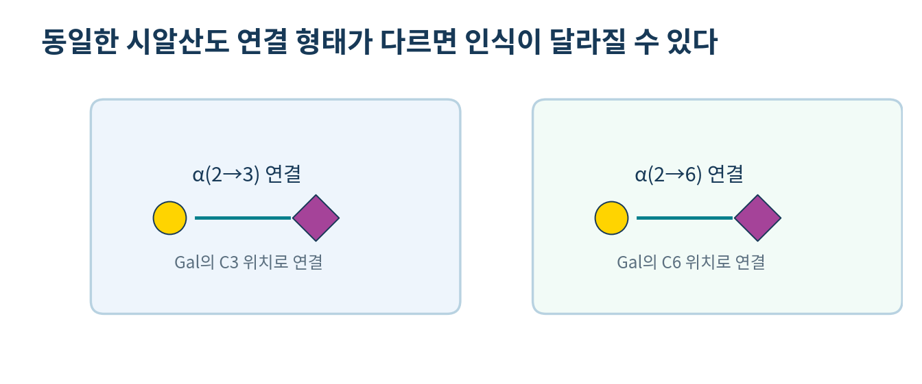
- Influenza haemagglutinin recognises sialylated glycans, with human-adapted lineages tending toward alpha2,6 and avian lineages toward alpha2,3 linkages
- Lineage and variant properties vary widely, so linkage preference alone does not fix host range
- Decoy molecules that mimic the receptor are under study, but binding in vitro does not guarantee a therapeutic effect
05
Glycomics methods and data interpretation
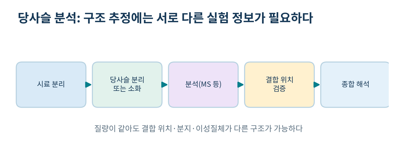
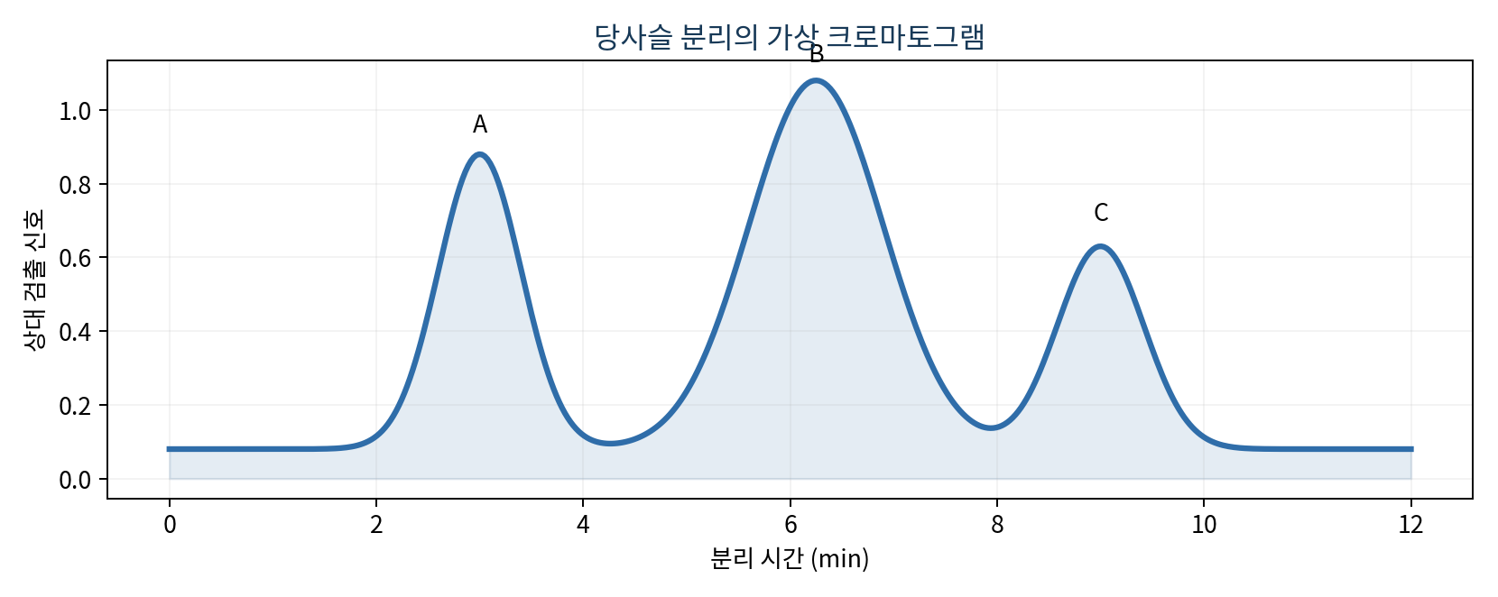
- Mass spectrometry gives molecular mass and fragment ions, and combining it with chromatography, specific glycosidases, labelling and linkage analysis strengthens the case
- Equal composition can still differ in branching and linkage, so equal mass does not fix structure
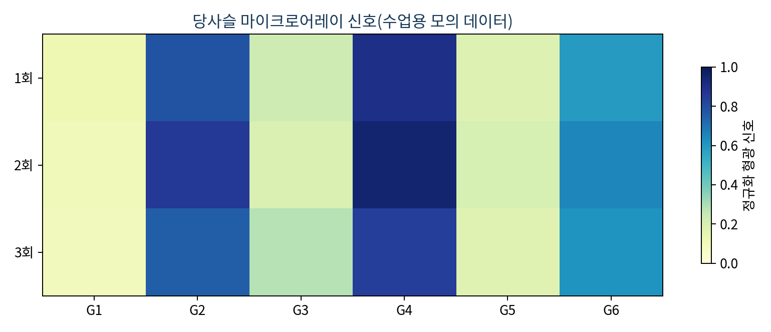
- Lectin microarrays immobilise lectins while glycan microarrays immobilise glycans, and both compare selective binding signals
- Either format requires control of background, non-specific binding, sample amount, labelling, concentration and layout
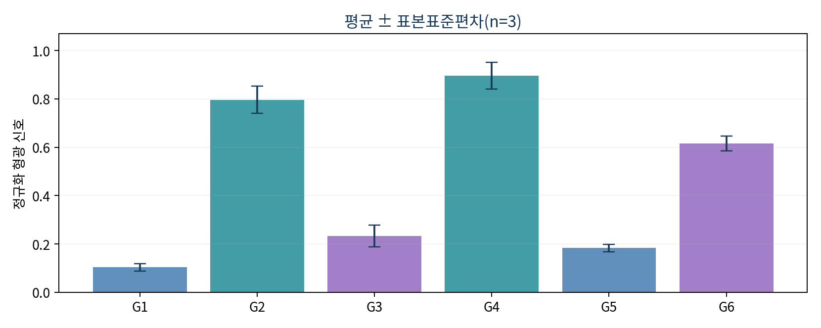
- Example: readings of 0.90, 0.95 and 0.84 for G4 give a mean of 0.897, and 0.78, 0.86 and 0.75 for G2 give a sample standard deviation of 0.057
- Reproducibility alone does not prove specificity, so negative controls and competition experiments are needed
- One chromatographic peak does not imply a single chemically pure structure
References
| Concept | Meaning | Example |
|---|---|---|
| Sugar composition | Identity and number of monosaccharides | Two GlcNAc and one Man |
| Linkage position | Which carbons are joined | alpha(1-3), beta(1-4) |
| Configuration | Alpha or beta at the anomeric carbon | alpha-D-Gal, beta-D-Gal |
| Branching | Extension of chains in several directions | Branches of an N-glycan |
| Terminal sugar | End most accessible for direct recognition | Fuc, Neu5Ac |
| Family | Typical recognition | Representative role |
|---|---|---|
| Selectin | Specific fucosylated and sialylated determinants | Leukocyte adhesion and migration |
| Galectin | Glycans containing beta-galactoside | Cell signalling, immunity, cell-cell interaction |
| Siglec | Sialic-acid-bearing glycans, specificity varying by protein | Immune cell regulation |
| Mannose 6-phosphate receptor | Mannose 6-phosphate on glycans | Sorting and transport of lysosomal enzymes |
| Condition | Purpose | Caution |
|---|---|---|
| Background, no sample | Confirms baseline label and substrate signal | Account for it properly in the raw data |
| Negative control glycan | Detects non-specific binding | Does not guarantee absolute negativity for every lectin |
| Known positive control | Verifies that the assay works | Control substrate concentration and labelling |
| Free sugar competition | Tests dependence on sugar recognition | Control solubility and non-specific effects |
| Technical replicate | Assesses measurement precision | Distinguish from biological replication |