01
DNA is a double helix that stores genetic information
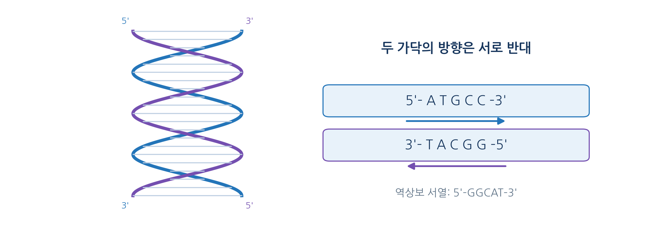
- The Watson-Crick duplex has two antiparallel strands with the backbone outside and the bases inside
- The complement of 5-prime ATGCC 3-prime is 3-prime TACGG 5-prime, written 5-prime GGCAT 3-prime
- That is the reverse complement, and confusing it with a simple complement causes errors in primer design and sequence analysis
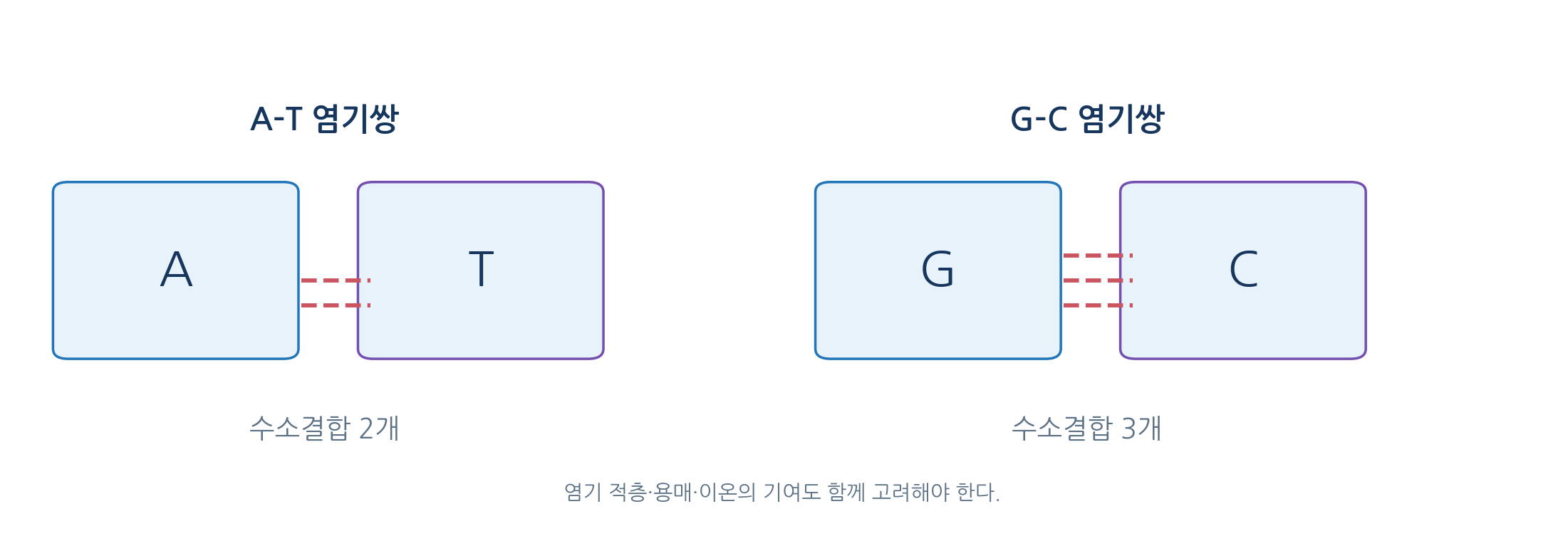
- A-T pairs have two hydrogen bonds and G-C pairs three, yet stacking, hydration, ions and neighbours also set stability
- Equal GC content with a different order can still change melting temperature and local shape
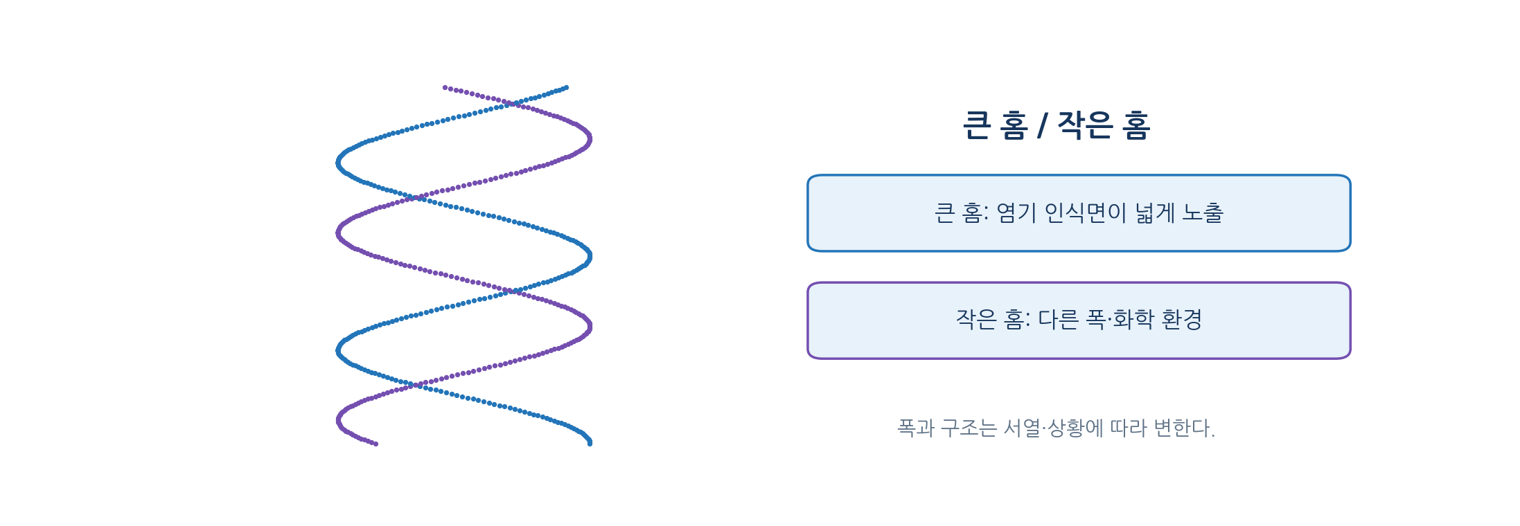
- B-DNA has a major and a minor groove of different width between the two backbones
- The grooves expose hydrogen-bond donors and acceptors and non-polar groups in different arrangements
- DNA-binding proteins read the chemical pattern together with local bending
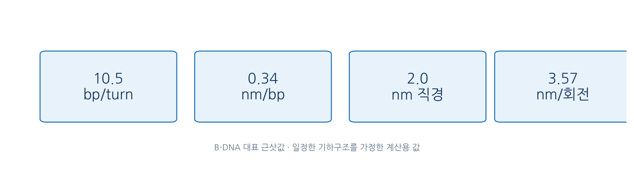
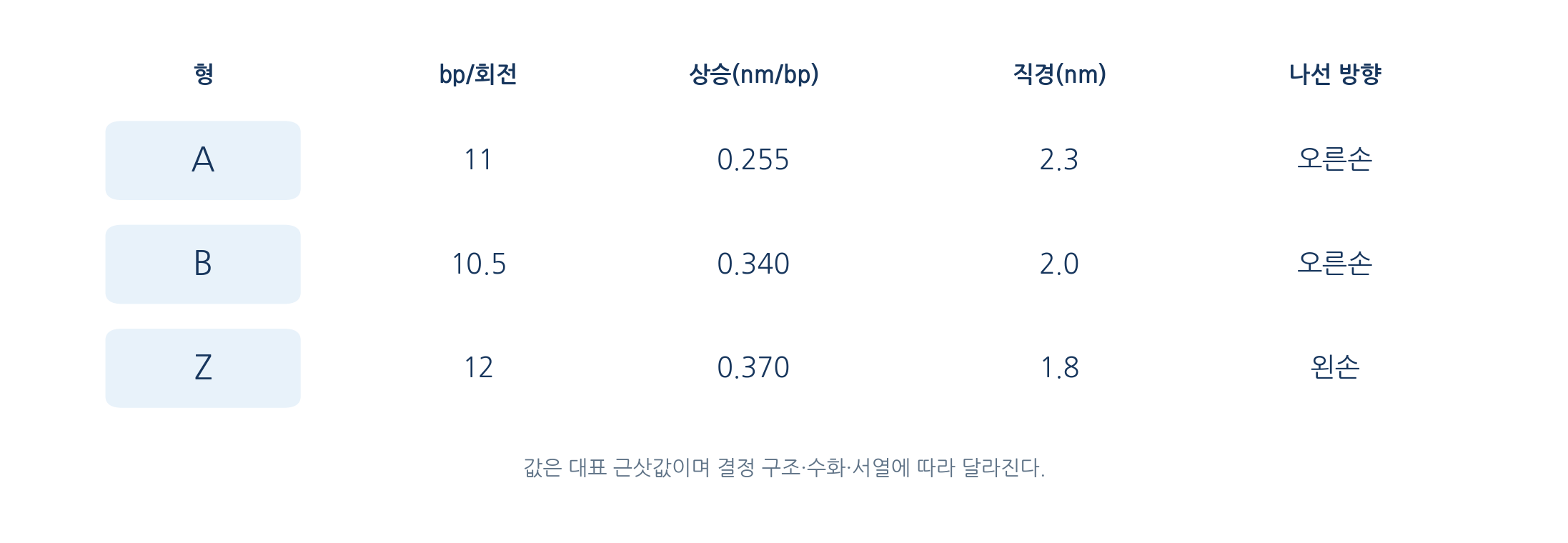
- Typical B-DNA values are a diameter of 2.0 nm, a rise of 0.34 nm per bp and 10.5 bp per turn, giving a pitch near 3.57 nm
- Real DNA bends and twists with sequence, hydration, tension and protein binding
- Example: 1,050 bp gives an axial length of 357 nm and 100 turns
02
DNA can adopt several three-dimensional forms
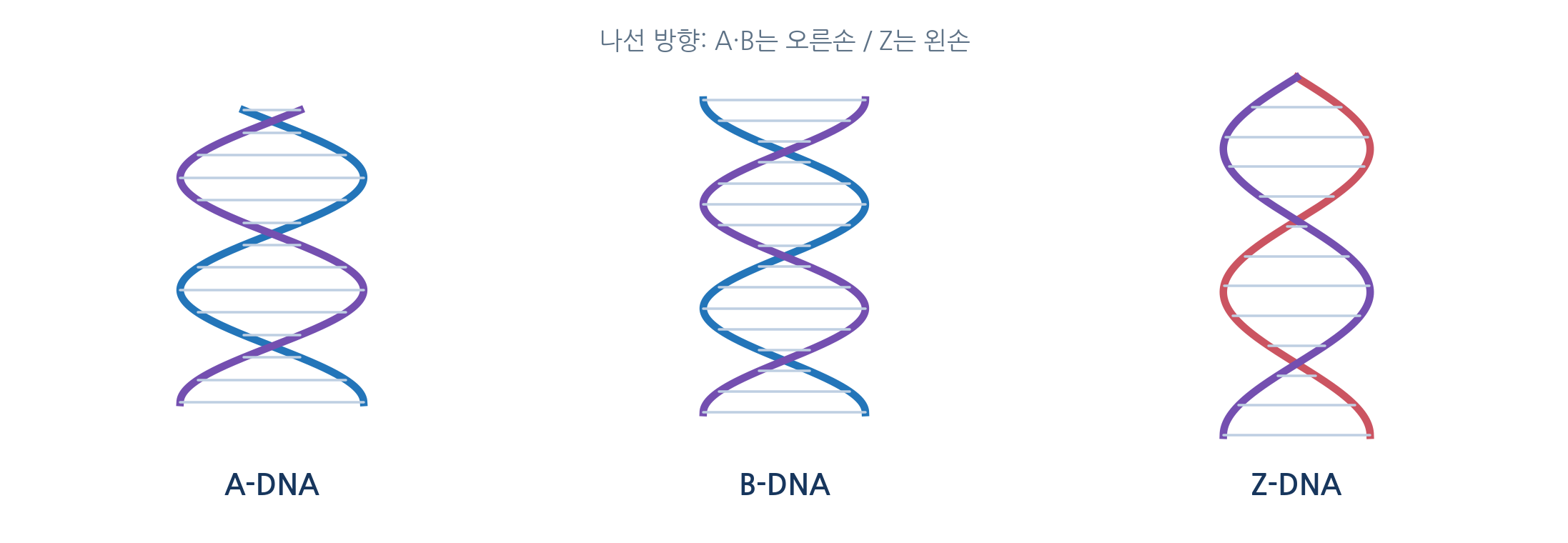
- A and B DNA are both right-handed, but A is shorter and wider with strongly tilted base pairs
- A-DNA appears at reduced hydration and in DNA-RNA duplexes, and double-stranded RNA also adopts an A-like form
- Z-DNA is a left-handed helix with a zigzag backbone, favoured by alternating purine-pyrimidine sequences
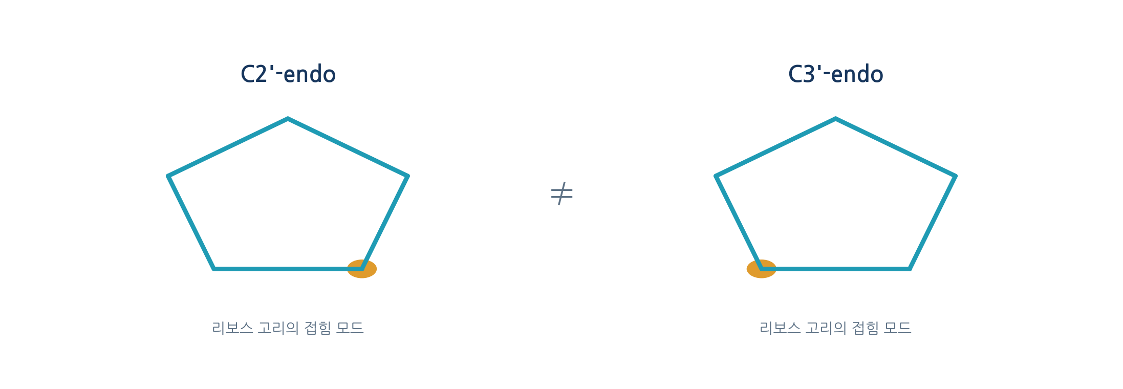
- The pentose ring is not flat but puckered, with B-DNA tending to C2-prime-endo and A-DNA and A-RNA to C3-prime-endo
- Sugar pucker constrains backbone geometry and base pair placement and so shapes the helix
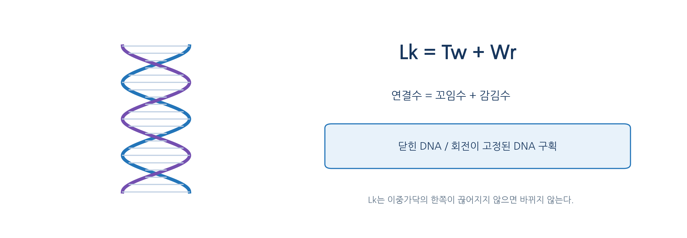
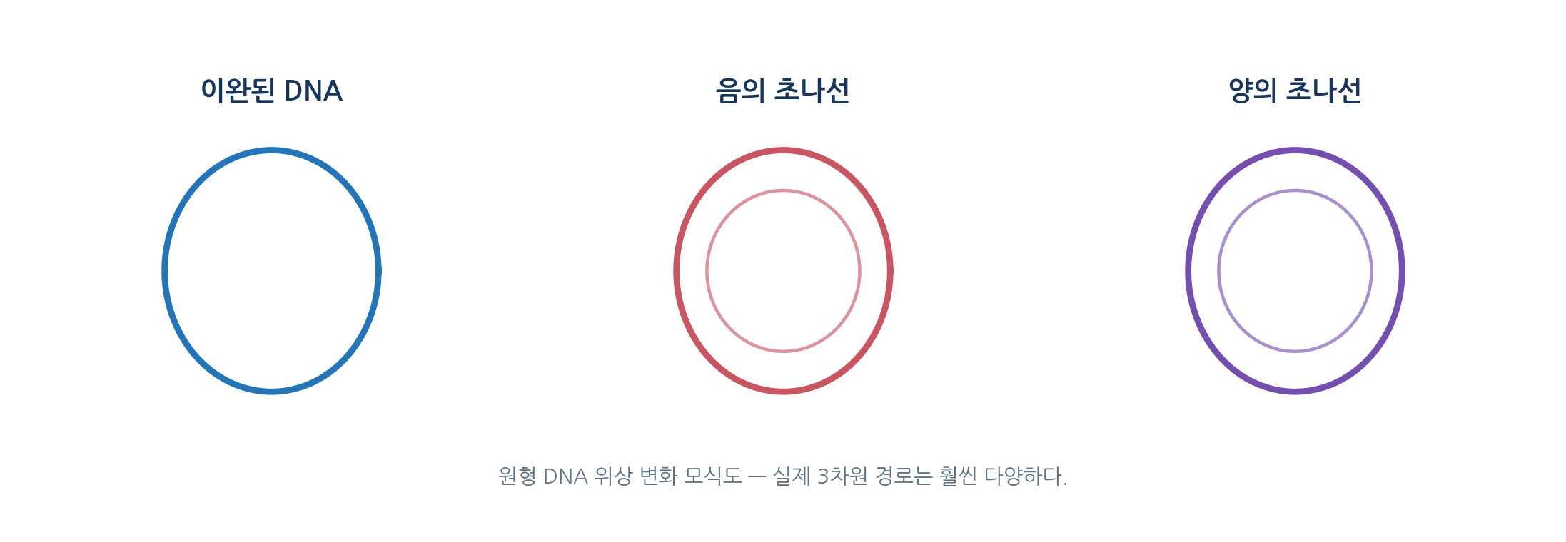
- Closed circular or rotationally fixed DNA is described by Lk = Tw + Wr
- Tw is local twist and Wr is the spatial coiling of the duplex axis
- Example: 2,100 bp with Lk0 near 200 and Lk 190 gives ΔLk of -10 and a superhelical density of -0.05, a negatively supercoiled state
- A nick that allows free rotation means Lk cannot be treated as a topological invariant
03
Certain sequences form unusual structures
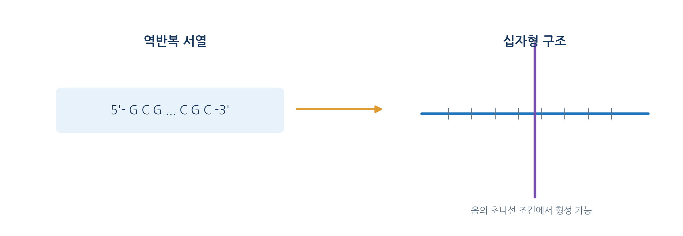
- Inverted repeats can form hairpins when strands separate and cruciforms in a closed duplex
- Formation depends on supercoiling, salt and surrounding conditions as well as sequence
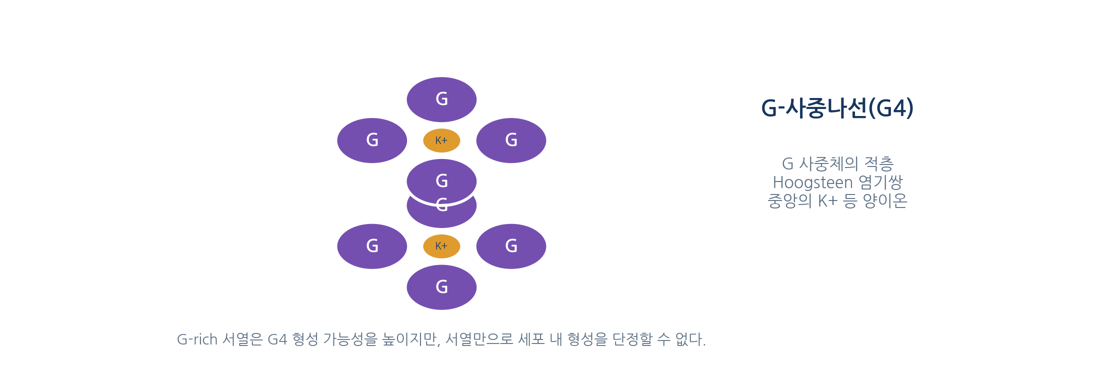
- A G-quartet is a planar arrangement of four guanines held by Hoogsteen hydrogen bonds, and stacked quartets give a G-quadruplex
- Monovalent cations such as K+ stabilise G4 in the central channel
- Formation capacity can be shown in vitro, but cellular occupancy and function are separate questions
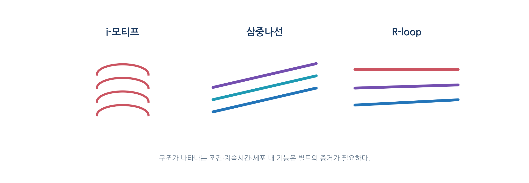
- Cytosine-rich sequences can form i-motifs built on C-C+ base pairs
- A triplex forms when a third strand binds the major groove of an existing duplex through Hoogsteen interactions
- An R-loop forms when RNA pairs with one DNA strand and displaces the other
- Capacity to form, observation in purified samples and a claim of cellular function require different levels of evidence
04
Messenger RNA encodes a polypeptide chain
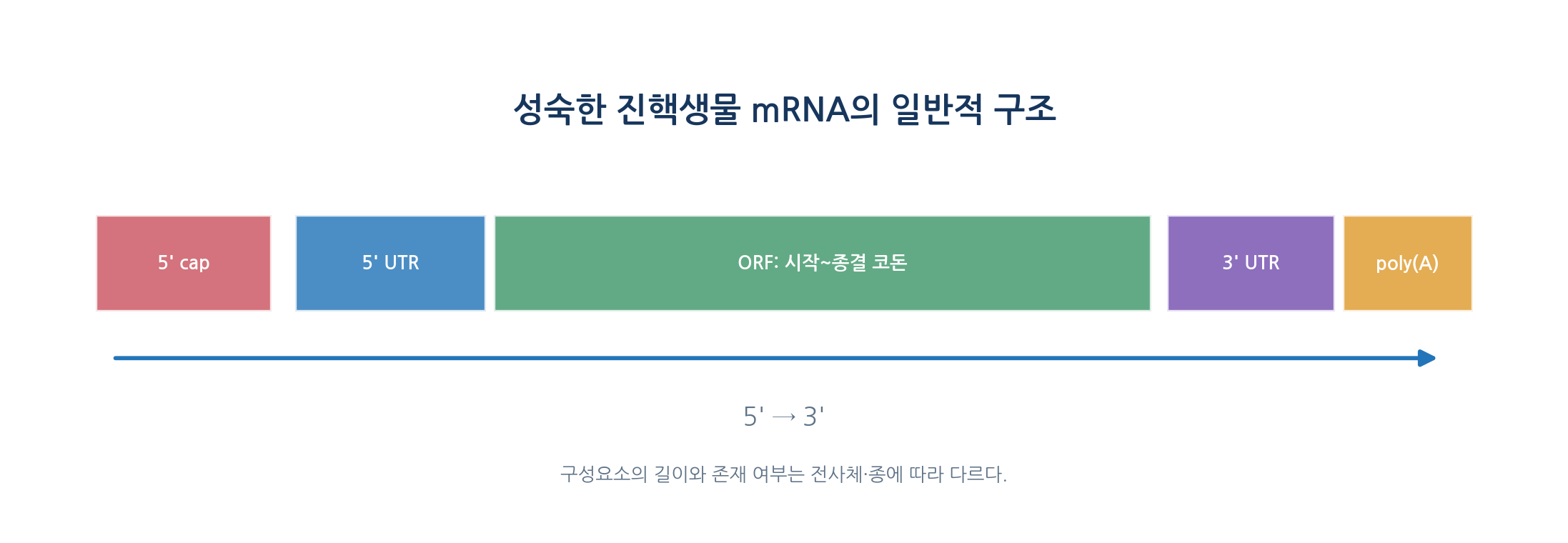
- mRNA supplies codons and comprises a 5-prime cap, 5-prime UTR, ORF, 3-prime UTR and poly(A) tail
- The ORF contains the start and stop codons, and the stop codon specifies no amino acid
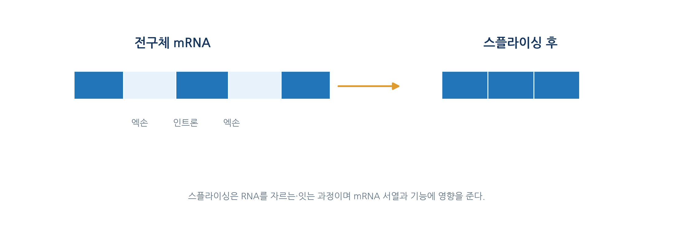
- Precursor mRNA matures through 5-prime capping, splicing, 3-prime cleavage and polyadenylation
- The cap has an unusual 5-prime to 5-prime triphosphate linkage and the poly(A) tail supports stability, translation and transport
- Alternative splice site choice yields several products from one gene
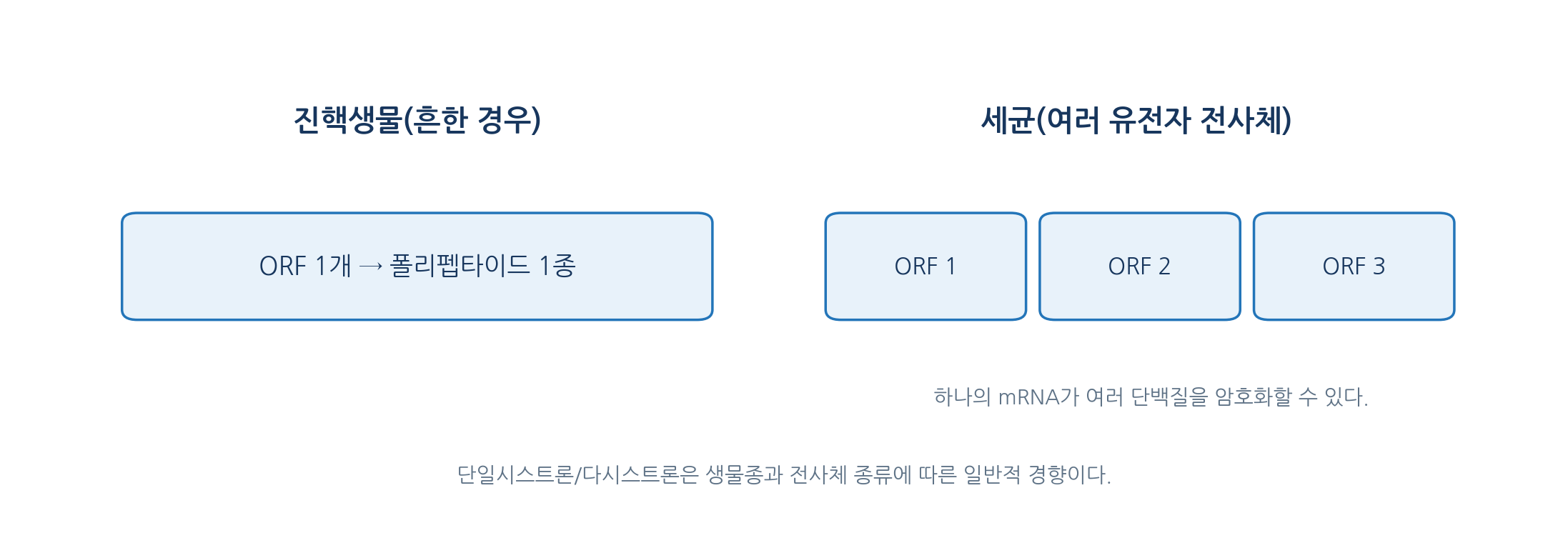
- Eukaryotic mRNA is usually monocistronic and bacterial mRNA often polycistronic, though exceptions exist
- Example: a 1,203 nucleotide ORF is 401 codons, giving 400 residues once the stop codon is excluded
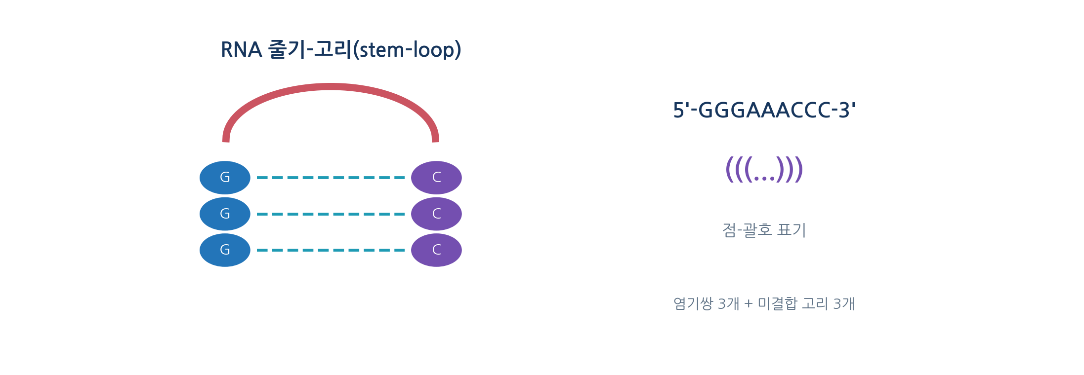
- A 5-prime UTR stem-loop changes initiation factor access, and 3-prime UTR structure couples to protein and regulatory RNA action
- The iron response element of ferritin mRNA binds iron-regulatory proteins
05
Many RNAs have complex three-dimensional structure
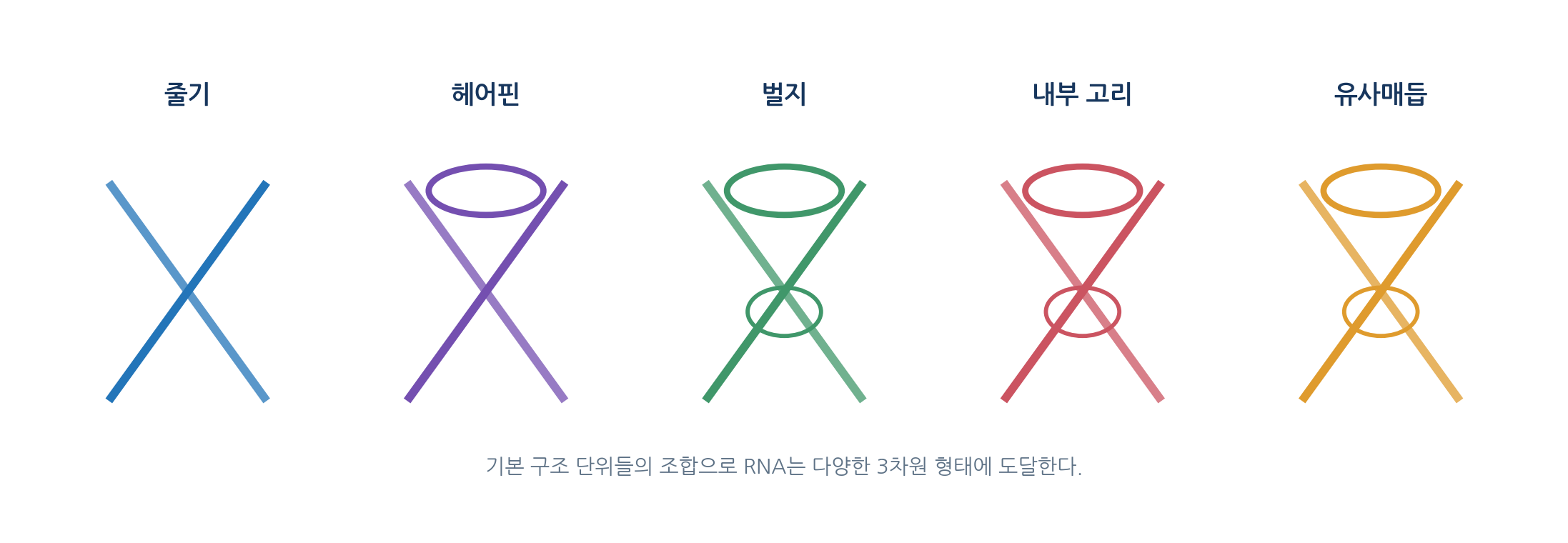
- Secondary structure is described with stems, hairpin loops, bulges, internal loops, multibranch loops and pseudoknots
- Tertiary structure adds long-range contacts, the arrangement of bases, sugars and phosphates, and metal and protein binding
- Besides A-U and G-C there are G-U wobble pairs and non-canonical interactions
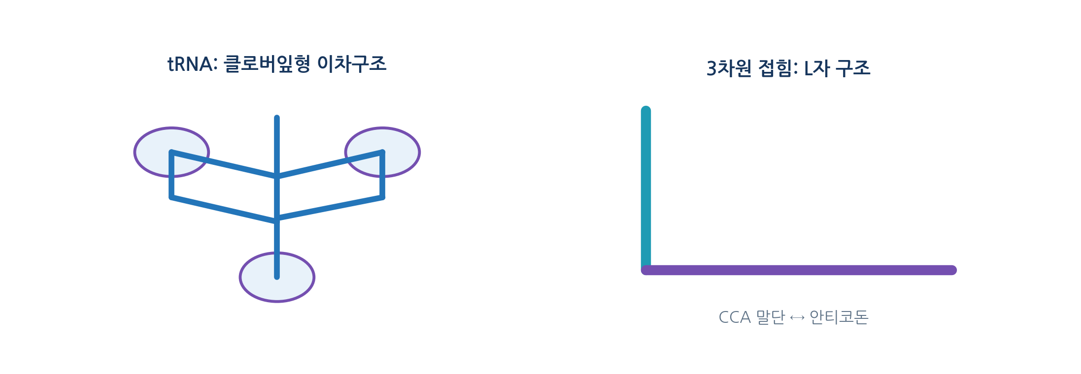
- tRNA has 70 to 90 nucleotides, drawn as a cloverleaf in two dimensions but folded into an L shape in three
- Amino acids attach to the 3-prime CCA end and the anticodon pairs with the codon
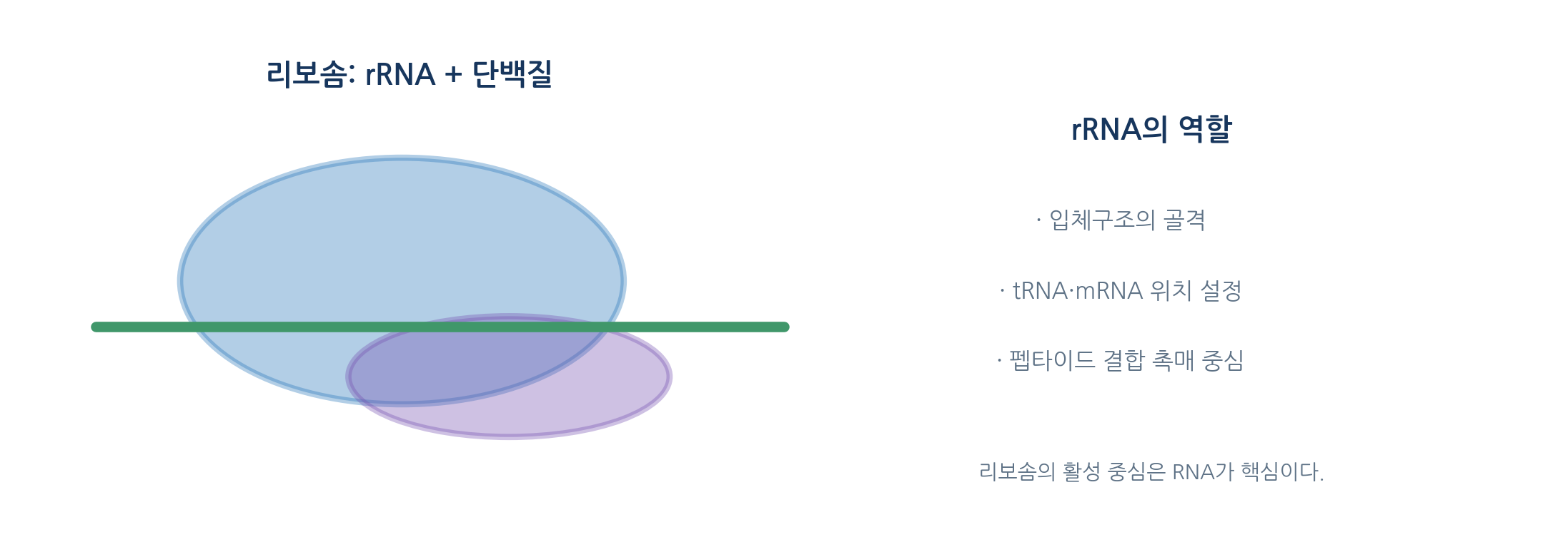
- In the ribosome, rRNA forms the scaffold, positions tRNA and mRNA, and provides the peptidyl transferase centre
- Ribozymes catalyse reactions themselves, and the ribosome is the leading biological example
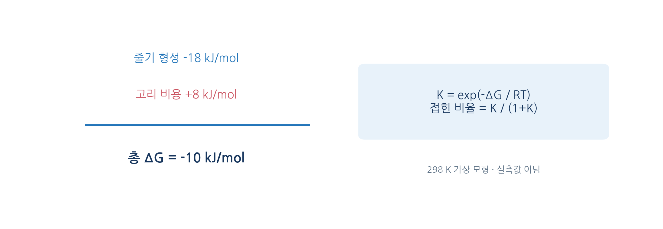
- Secondary structure prediction uses nearest-neighbour free energies that count adjacent pairs and loop types
- Example: ΔG° of -10.0 kJ/mol gives K near 56.5 and a folded fraction of about 98.3 percent
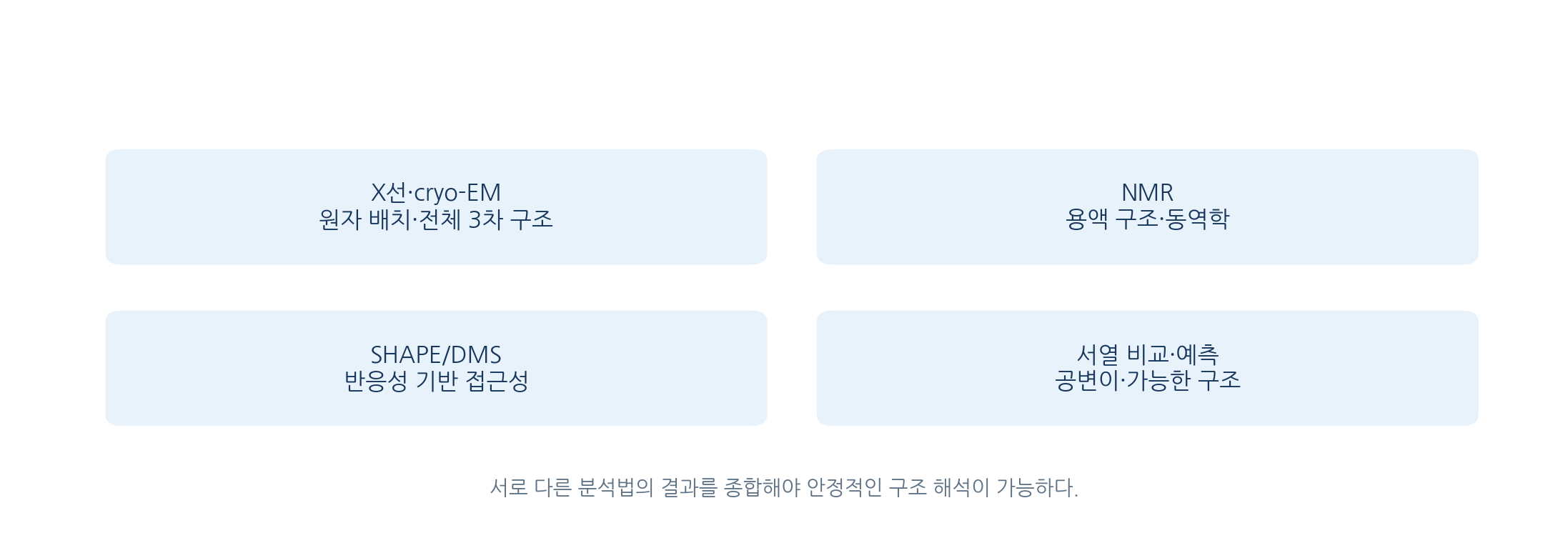
- X-ray, cryo-EM, NMR, SHAPE, DMS, covariation analysis and prediction are complementary
References
| Feature | A-DNA | B-DNA | Z-DNA |
|---|---|---|---|
| Helix sense | Right-handed | Right-handed | Left-handed |
| Base pairs per turn | About 11 | About 10.5 | About 12 |
| Rise per bp in nm | About 0.255 | About 0.340 | About 0.370 |
| Diameter in nm | About 2.3 | About 2.0 | About 1.8 |
| Character | Wide and short with strongly tilted pairs | The usual form in aqueous solution | Zigzag backbone favoured by alternating sequences |
| Observation | What it supports | Further evidence needed |
|---|---|---|
| G-rich sequence | Possible G4 formation | Structure-specific assays and cellular detection |
| Change in CD spectrum | Possibly an average structural change | Independent structural analysis and controls |
| Signal from a specific antibody | The structure or antigen may be present | Specificity and cross-reactivity testing |
| X-ray, NMR or cryo-EM result | Three-dimensional constraints under those conditions | Cellular dynamics and physiological abundance |